

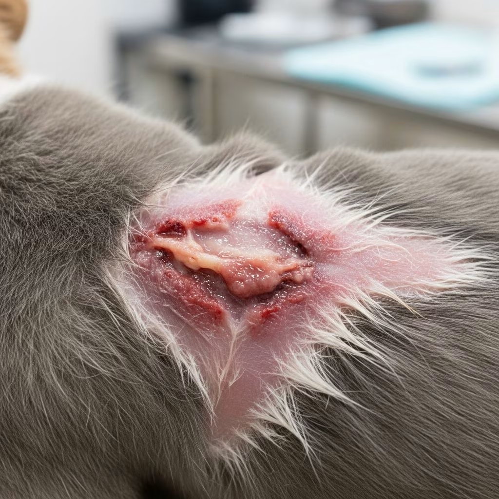
Effective wound management in small animals such as dogs and cats relies on understanding the natural healing process and applying targeted interventions to support it. This guide details the biological stages, immediate care protocols, cleaning methods, tissue removal strategies, repair techniques, and protective measures to promote swift and complication-free recovery.
The Biological Journey of Wound Repair
Wound healing in small pets unfolds through distinct overlapping phases that restore tissue integrity. Immediately after injury, the inflammatory phase activates as blood vessels constrict then dilate, forming a clot to staunch bleeding and ward off pathogens. Neutrophils and macrophages flood the site, clearing bacteria and dead cells through phagocytosis.
Transitioning next, the proliferative phase builds new tissue. Fibroblasts produce collagen, forming granulation tissue—a pink, vascular bed that fills the defect. Epithelial cells migrate across the surface, while angiogenesis creates fresh blood supply. Contraction pulls wound edges together via myofibroblasts, reducing the healing area.
Finally, the remodeling phase refines the scar over months, with collagen realignment boosting tensile strength up to 80% of original tissue. Disruptions like infection or poor nutrition can stall these steps, prolonging recovery.
- Inflammatory phase: Hours to days; hemostasis and debris cleanup.
- Proliferative phase: Days to weeks; tissue formation and contraction.
- Remodeling phase: Weeks to years; scar maturation.
Prioritizing Patient Stability Before Wound Care
Before addressing any injury, evaluate the pet’s overall condition. Signs of shock, such as pale gums, rapid pulse, or lethargy, demand immediate stabilization via fluids, oxygen, and monitoring. Open wounds often mask internal issues like diaphragmatic hernias or pneumothorax.
Apply gentle pressure with clean gauze to control bleeding, avoiding tourniquets which risk tissue damage. Cover the wound loosely with a sterile dressing to shield from dirt while awaiting professional help. Analgesics like opioids provide comfort without masking vital assessments.
| Stability Check | Actions |
|---|---|
| Airway/Breathing | Ensure clear passage; supplement oxygen if needed. |
| Circulation | IV fluids for hypotension; control external hemorrhage. |
| Neurologic Status | Assess consciousness; manage pain. |
Cleaning Wounds: Lavage Techniques for Purity
Thorough irrigation removes contaminants, slashing infection risk. Use copious sterile saline (at least 1-2 liters for large wounds) delivered via syringe or pressure device at 8-12 psi to dislodge debris without traumatizing viable tissue. Add antiseptics like dilute chlorhexidine if heavy contamination exists, but avoid cytotoxics on clean wounds.
Lavage enhances visibility for deeper inspection, revealing hidden foreign bodies or tendon damage. Perform under sedation to minimize stress. Collect swab samples from the wound base pre-lavage for culture if infection suspected, guiding antibiotic choice.
- Prepare: Clip fur widely, don sterile gloves.
- Irrigate: High volume, moderate pressure.
- Inspect: Probe gently for extent.
Debridement: Excising the Dead to Save the Living
Non-viable tissue harbors bacteria and impedes healing, necessitating prompt removal. Sharp debridement with scalpel or scissors targets devitalized areas, preserving healthy margins. For selective work, use curettes on granulation beds.
Autolytic methods employ occlusive dressings to harness enzymes from the pet’s cells. Mechanical wet-to-dry gauze pulls debris on changes, ideal initially but harsher. Enzymatic gels with collagenase aid chronic cases. Repeat as needed until bleeding healthy tissue appears.
| Method | Best For | Pros/Cons |
|---|---|---|
| Sharp | Acute, necrotic edges | Precise; requires anesthesia. |
| Autolytic | Clean, low-exudate | Gentle; slower. |
| Mechanical | Sloughy wounds | Effective debridement; painful. |
Closure Strategies: Sealing Wounds for Swift Recovery
Primary closure suits clean, fresh lacerations under 6-12 hours old post-debridement. Apposition with absorbable subcutaneous sutures eliminates dead space, topped by skin staples or nylon for strength. Tension-relieving patterns like walking sutures suit high-movement areas.
Delayed secondary closure follows 3-5 days of open care for contaminated injuries, leveraging early granulation. Ensure edges are viable before suturing. Not all wounds close; extensive defects demand grafts or flaps.
- Clean <6h: Primary closure.
- Infected: Open 24-72h, then secondary.
- Large gaps: Second intention or reconstructive surgery.
Open Management: Harnessing Natural Healing Forces
When closure risks failure, second-intention healing fills defects via granulation, contraction, and epithelization. Ideal for bites, deglovings, or abscesses. Regular debridement and bandaging prevent desiccation and infection.
Moist environments accelerate progress over dry scabbing. Honey dressings draw exudate while antimicrobial; foams absorb heavy drainage; hydrogels hydrate eschar.
Bandaging Mastery: Protection and Promotion
Bandages maintain moisture, absorb fluids, immobilize, and protect. Layers include non-adherent primary contact, absorbent secondary, cast-padding tertiary, and outer conforming wrap. Change every 1-3 days, assessing progress.
Complications like strikes or slippage require E-collars and checks. Tailor to site: Robert Jones for limbs, tie-over for trunks.
| Layer | Material | Function |
|---|---|---|
| Primary | Non-stick pads | Contact, debride gently. |
| Secondary | Gauze, foams | Absorb exudate. |
| Tertiary | Cast padding | Cushion, conform. |
| Outer | Elastikon | Secure, waterproof. |
Factors Influencing Healing Success
Age, nutrition, comorbidities like diabetes, and steroids hinder progress. Protein-calorie deficits stall collagen synthesis; zinc aids epithelialization. Minimize tension and motion. Broad-spectrum antibiotics cover empiric therapy pending cultures.
- Systemic support: Balanced diet, pain control.
- Avoid: Smoking exposure (owners), excessive steroids.
Advanced Therapies for Challenging Wounds
Chronic non-healers benefit from negative pressure therapy, pulling exudate while stimulating granulation. Biologic scaffolds like xenografts provide matrix. Laser or ultrasound boosts circulation. Veterinary platelet-rich plasma delivers growth factors.
Monitor via photos and measurements for objective tracking.
Common Pitfalls and Prevention Tips
- Over-bandaging: Causes maceration; ensure breathability.
- Rushed closure: Breeds dehiscence; debride thoroughly.
- Inadequate lavage: perpetuates infection; use volumes liberally.
- Pain neglect: Stress delays healing; multimodal analgesia.
FAQs on Small Animal Wound Care
What is the first step after finding a wound on my pet?
Stabilize the animal, control bleeding with pressure, cover loosely, and seek veterinary care promptly.
How often should bandages be changed?
Every 1-3 days or if soiled/striking, under professional guidance to avoid trauma.
Can all wounds be sutured immediately?
No; contaminated or old wounds require open management first to prevent abscesses.
What home care supports healing?
Prevent licking/chewing with collars, ensure rest, provide nutritious food, and follow vet instructions.
When does a wound need emergency attention?
If deep, bleeding profusely, near joints/eyes, or accompanied by lethargy/fever.
Monitoring Progress for Optimal Outcomes
Weekly reassessments gauge contraction, granulation quality, and exudate. Healthy progress shows shrinking pink beds with advancing epithelium. Stagnation prompts culture, biopsy, or therapy shifts. Patient education empowers owners for compliance.
References
- Initial Wound Management in Small Animals — MSD Veterinary Manual. 2023. https://www.msdvetmanual.com/emergency-medicine-and-critical-care/wound-management-in-small-animals/initial-wound-management-in-small-animals
- Principles of Wound Management and Wound Healing in Exotic Pets — PMC/NCBI. 2015-10-01. https://pmc.ncbi.nlm.nih.gov/articles/PMC4663678/
- Basic Principles of Wound Management (Proceedings) — dvm360. 2022. https://www.dvm360.com/view/basic-principles-wound-management-proceedings
- Principles of Wound Care & Bandaging Techniques — Today’s Veterinary Practice. 2023. https://todaysveterinarypractice.com/wound-management/todays-technician-principles-of-wound-care-bandaging-techniques/
- Basic Principles of Wound Care — Today’s Veterinary Nurse (PDF). 2020-03. https://todaysveterinarynurse.com/wp-content/uploads/sites/3/2020/03/Jaeger_WoundCare_TVNSpring2020.pdf



