

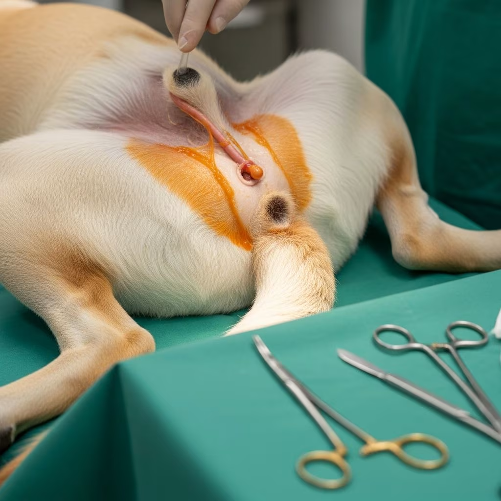
Congenital urethral anomalies represent a diverse group of structural abnormalities in the urinary tract that can significantly impact animal health, particularly in young patients. These defects, ranging from incomplete development to misplaced openings, often manifest early in life and require prompt veterinary intervention to prevent complications like infections or kidney damage.
Understanding the Basics of Urethral Development
The urethra forms during embryonic development as a critical conduit for urine expulsion. Disruptions in this process can lead to various malformations. In veterinary practice, these issues are more prevalent in certain breeds and species, influenced by genetic and environmental factors. Early detection through routine exams is key to managing long-term prognosis.
Common Types of Urethral Anomalies
Several distinct urethral defects occur in animals, each with unique anatomical features and clinical implications.
- Hypospadias: This condition involves the urethral opening positioned abnormally on the ventral penis surface, leading to spraying urination and potential fertility issues in males.
- Urethral Duplication: Rare cases feature two urethral channels, one normal and one ectopic, often causing urine leakage from unexpected sites like near the anus.
- Urethral Prolapse: Mucosal tissue protrudes through the urethral orifice, commonly seen in young intact male dogs of specific breeds.
- Urethral Atresia or Stenosis: Complete or partial blockages hinder urine flow, resulting in severe distension upstream.
- Fistulas: Abnormal connections between urethra and rectum or skin allow urine diversion, predisposing to infections.
Species-Specific Prevalence and Presentations
Urethral anomalies vary by animal type, with dogs showing the highest incidence among companion animals.
| Species | Common Anomalies | Predisposed Groups | Typical Age of Onset |
|---|---|---|---|
| Dogs | Duplication, prolapse, hypospadias | Males, brachycephalics, Yorkshire terriers | Birth to 2 years |
| Cats | Hypoplasia, sphincter incompetence | Females, young kittens | Neonatal period |
| Horses | Associated ectopias | Fillies | Early foalhood |
| Livestock (cattle, sheep) | Atresia, fistulas | Males | Birth |
Clinical Signs and Complications
Owners often notice abnormal urination patterns first. Key symptoms include:
- Urine dribbling from non-standard locations.
- Frequent urinary tract infections due to stagnant urine.
- Straining or pain during voiding.
- Swelling in genital or perineal areas.
- Incontinence, especially post-exercise or at rest.
Untreated cases risk hydronephrosis, renal failure, or chronic dermatitis from urine scalding.
Diagnostic Approaches
Accurate diagnosis combines history, physical exams, and imaging.
- Physical Examination: Reveals ectopic openings or prolapsed tissue.
- Ultrasound: Assesses urethral diameter, bladder wall, and kidney status.
- Contrast Studies: Retrograde urethrography or cystography maps anomalous paths.
- CT/MRI: Provides detailed 3D views for complex duplications.
- Cystoscopy: Direct visualization confirms internal defects.
Lab tests like urinalysis detect infections or crystals early.
Treatment Strategies: Medical and Surgical
Management depends on anomaly severity and patient stability. Medical options stabilize before surgery.
Medical Management
Antibiotics treat infections, while alpha-agonists or estrogen supplements aid sphincter tone in incompetence cases. Bulking agents injected endoscopically narrow the lumen, improving continence.
Surgical Interventions
Surgery is definitive for most defects.
- For Prolapse: Resection of protruded mucosa with purse-string suturing.
- Duplication Correction: Perineal or abdominal approaches excise ectopic segments, preserving main channel patency.
- Hypospadias Repair: Urethroplasty reconstructs normal opening.
- Fistula Closure: Layered suturing separates tracts.
Post-op care includes catheterization, antibiotics, and monitoring for strictures.
Case Studies Highlighting Challenges
A 2-year-old Yorkshire terrier presented with dual urine streams from urethra and anus, diagnosed via CT as perineal urethral duplication with cryptorchidism. Initial perineal surgery left a remnant causing obstruction; a second abdominopelvic procedure resolved it successfully.
In kittens, congenital strictures lead to acute retention, requiring urgent perineal urethrostomy for relief.
Prognosis and Long-Term Care
Success rates exceed 80% with timely intervention, though recurrences or incontinence persist in 20-30% of cases. Regular follow-ups monitor function via imaging and cultures. Owners should watch for straining or blood in urine.
Preventive Measures and Genetic Considerations
Breeding from affected lines is discouraged. Genetic screening in predisposed breeds like bulldogs aids prevention. Neutering post-correction reduces prolapse risk.
FAQs
What causes urethral anomalies in pets?
Multifactorial origins include genetic mutations and developmental errors during embryogenesis.
Can urethral prolapse resolve without surgery?
Mild cases in young dogs may regress with medical therapy, but severe ones need resection.
Is incontinence common after urethral surgery?
Possible in 10-20% of cases; managed with medications or additional procedures like colposuspension.
How do I know if my puppy has a urethral defect?
Observe urination: spraying, dribbling, or straining signals a vet visit.
Are these anomalies heritable?
Yes, particularly in certain dog breeds; avoid breeding affected animals.
Advancements in minimally invasive techniques promise improved recovery times and outcomes for animals with these challenging conditions.
References
- Case report: Post-surgical complication in a case of urethral duplication in a dog — Frontiers in Veterinary Science. 2022-10-11. https://www.frontiersin.org/journals/veterinary-science/articles/10.3389/fvets.2022.1013270/full
- Ureteral Anomalies in Animals — MSD Veterinary Manual. 2023. https://www.msdvetmanual.com/urinary-system/congenital-and-inherited-anomalies-of-the-urinary-system/ureteral-anomalies-in-animals
- Urethral Incompetence in Dogs: Updates in Management — Today’s Veterinary Practice. 2022. https://todaysveterinarypractice.com/urology-renal-medicine/canine-urethral-incontinence/
- A rare case of a complex of multiple congenital anomalies including hypospadias, atresia ani and urethrorectal fistula in a dog — Veterinarni Medicina. 2018-01-08. http://vetmed.agriculturejournals.cz/pdfs/vet/2018/01/08.pdf
- Urethral Prolapse — Dallas Veterinary Surgical Center. 2023. https://www.dvsc.com/urethral-prolapse



