

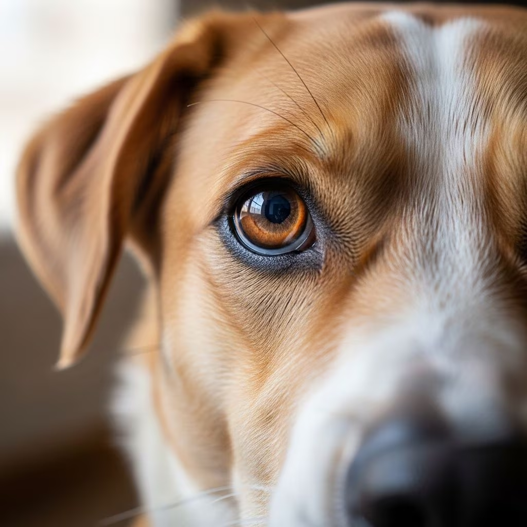
Observing one of your dog’s pupils larger than the other can be alarming for any pet owner. This condition, known as anisocoria, occurs when the pupils— the black centers of the eyes—fail to match in size. In healthy dogs, pupils adjust symmetrically to light changes, but asymmetry signals potential issues ranging from minor irritations to life-threatening disorders. Understanding the anatomy behind this and recognizing when it’s an emergency is crucial for timely intervention.
Understanding Canine Eye Anatomy
Dogs possess sophisticated eye structures adapted for their predatory heritage, enabling sharp motion detection and low-light vision. Key components include the cornea, a clear front layer that protects and refracts light; the iris, the colored ring controlling light entry; and the pupil, the central opening that dilates or constricts accordingly. Behind these lie the lens for focusing light onto the retina, and the sclera, the white outer layer. Dogs also feature a third eyelid for extra protection and moisture, plus a tapetum lucidum—a reflective layer boosting night vision.
Normally, both pupils react identically: enlarging in dim conditions to capture more light and shrinking in brightness via iris muscles. The Merck Veterinary Manual explains that the iris’s sphincter muscle constricts the pupil in light, while dilator muscles expand it in darkness, ensuring balanced vision. Disruptions here lead to unequal sizes.
What Triggers Unequal Pupil Sizes?
Anisocoria stems from imbalances in the nervous system regulating pupil response or direct eye damage. It divides into benign and pathological categories.
Benign or Physiological Causes
- Lighting Differences: Brief asymmetry from uneven light exposure on each eye, resolving quickly.
- Third Eyelid Influence: The nictitating membrane partially covering one eye can mimic pupil changes.
- Minor Irritations: Temporary responses to dust or allergens causing localized reactions.
These often self-correct without intervention.
Neurological Factors
The brain and nerves dictate pupil control. Issues like Horner’s Syndrome damage sympathetic nerves, constricting one pupil (miosis) and sometimes causing a droopy eyelid or sunken eye. Parasympathetic overactivity from oculomotor nerve problems enlarges one pupil (mydriasis). Head trauma, brain tumors, or infections can provoke this.
Eye-Specific Problems
| Condition | Pupil Effect | Associated Signs |
|---|---|---|
| Glaucoma | Dilated, unresponsive | Eye redness, pain, cloudiness, bulging |
| Uveitis | Constricted or irregular | Inflammation, squinting, tearing |
| Cataracts/Lens Luxation | Variable dilation | Cloudy lens, vision loss |
| Corneal Injury | One-sided constriction | Ulcer, blue haze, rubbing |
Glaucoma, with elevated intraocular pressure, demands immediate care to prevent blindness.
Breed Predispositions and Age Factors
Certain breeds like Cocker Spaniels, Basset Hounds, and Shih Tzus face higher glaucoma risks due to shallow eye sockets. Puppies’ eyes develop uniquely, with initial blue irises maturing later, but persistent anisocoria warrants checks. Senior dogs may show changes from degeneration.
Recognizing Warning Signs
Beyond size mismatch, watch for:
- Light insensitivity in the affected pupil.
- Redness, swelling, or discharge.
- Pawing at eyes, squinting, or head tilting.
- Behavioral shifts: lethargy, appetite loss, or aggression from pain.
- Cloudy corneas or vision impairment clues like bumping objects.
Acute onset with pain signals emergencies like glaucoma; chronic cases might indicate tumors.
When to Contact a Veterinarian
Don’t delay if anisocoria persists over minutes, accompanies pain, or affects vision. Even benign cases merit evaluation to exclude serious pathology. Vets use tools like fluorescein staining for ulcers, tonometry for pressure, and pupil response tests. Urgent care prevents irreversible damage.
Diagnostic Approaches
Exams start with history and observation, progressing to:
- Ophthalmoscopy: Magnified internal view.
- Tonometry: Pressure measurement; normal is 10-25 mmHg.
- Schirmer Tear Test: Dryness assessment.
- Neurological Evaluation: Reflex checks.
- Imaging: Ultrasound or CT for deeper issues.
These pinpoint causes accurately.
Treatment Options by Cause
Tailored therapies include:
- Medications: Eye drops for inflammation, pressure reducers for glaucoma.
- Surgery: Lens repair, tumor removal, or enucleation in severe cases.
- Supportive Care: Antibiotics for infections, pain relief.
Prognosis varies; early uveitis responds well, while advanced glaucoma may blind eyes.
Preventive Strategies for Eye Health
Proactive steps reduce risks:
- Regular vet checkups, especially for at-risk breeds.
- Avoid trauma: supervise play, use goggles for rough activities.
- Monitor for irritants; rinse eyes post-outdoors.
- Balanced diet supporting ocular health.
- Control systemic diseases like diabetes.
Genetic screening aids breeders.
FAQs on Dog Pupil Issues
Is anisocoria always serious in dogs?
Not always; physiological cases resolve alone, but veterinary confirmation is advised.
Can stress cause unequal pupils?
Yes, via nerve effects, but persistent cases need checks.
How quickly does glaucoma progress?
Rapidly; hours can lead to permanent damage.
Do puppies outgrow anisocoria?
Developmental fixes occur, but consult a vet.
What’s the home first aid for eye injury?
Gently flush with saline; seek professional care immediately.
Long-Term Management
Chronic conditions require lifelong monitoring, medications, or vision aids. Owners adapt environments for blind pets, using scents and routines. Support groups offer tips.
In summary, unequal pupils signal varied concerns in dogs. Swift action preserves vision and comfort. Consult professionals for personalized guidance.
References
- Dogs Eye Anatomy: Everything You Need To Know About Them — Zigzag.dog. 2023. https://zigzag.dog/en-us/blog/puppy-health/new-puppy-health/dog-eye-anatomy/
- Eye Structure and Function in Dogs — Merck Veterinary Manual. 2024-01-15. https://www.merckvetmanual.com/dog-owners/eye-disorders-of-dogs/eye-structure-and-function-in-dogs
- What Do Our Pets See? — ASPCA Pet Health Insurance. 2023. https://www.aspcapetinsurance.com/resources/seeing-the-world-through-our-pets-eyes/
- Dog Eye Structure: Anatomy & Functions — MP Labo. 2024. https://www.mplabo.com/en/questions/the-structure-of-the-eye/
- Dog Vision and Eye Anatomy: How Dogs See — PetMD. 2025-06-10. https://www.petmd.com/dog/general-health/how-do-dogs-see-world
- Structure of the Canine Eye — Whole Dog Journal. 2023-11-20. https://www.whole-dog-journal.com/health/structure-of-the-canine-eye/
- Understanding the Anatomy of the Eye (Cats and Dogs) — Dechra UK. 2024. https://www.dechra.co.uk/companion-animal/ophthalmology/anatomy-of-the-eye



