

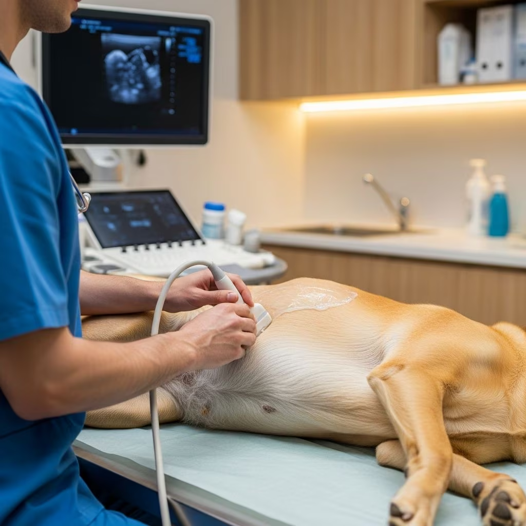
Understanding Ultrasound Examination in Dogs
An ultrasound examination, also known as ultrasonography, is a non-invasive imaging technique that allows veterinarians to visualize internal body structures by recording echoes of sound waves. This diagnostic tool has become essential in modern veterinary medicine, enabling practitioners to examine organs, tissues, and blood vessels without surgical intervention. Unlike X-rays that use radiation, ultrasound employs harmless sound waves to create real-time images of your dog’s internal organs, making it an exceptionally safe and effective diagnostic method for identifying health issues early.
How Veterinary Ultrasound Works
The ultrasound machine functions by emitting high-frequency sound waves through a handheld device called a transducer. These sound waves travel through your dog’s body and bounce off internal structures, creating live images that appear on a monitor. The transducer detects these returning echoes and converts them into detailed visual representations of organs, tissues, and blood flow patterns. This real-time imaging capability allows veterinarians to observe how organs are functioning at the moment of examination, providing immediate insights into your pet’s health status.
To enhance image quality and ensure accurate diagnosis, a water-based gel is applied to your dog’s skin before the transducer is placed. This gel provides optimal contact between the probe and skin, allowing sound waves to transmit effectively into the body without distortion caused by air gaps. The process is completely painless and non-invasive, making it suitable for dogs of all ages and health conditions.
Common Types of Ultrasound Examinations for Dogs
Veterinarians utilize various ultrasound techniques depending on the diagnostic needs of your dog. Understanding these different types can help you better comprehend what your veterinarian recommends:
Abdominal Ultrasound
The abdominal ultrasound is one of the most frequently performed examinations. It can search the abdomen for abnormalities or be performed on an emergency basis to detect life-threatening conditions such as internal bleeding or intestinal blockages. This comprehensive scan evaluates the liver, kidneys, spleen, stomach, intestines, and other abdominal structures, providing a complete picture of your dog’s digestive and urinary systems.
Echocardiogram
An echocardiogram is a specialized ultrasound examination of the heart that evaluates the heart chambers, blood flow through the heart, and the pericardial sac that surrounds the heart. This diagnostic test is the standard procedure for diagnosing and monitoring heart disease in dogs, allowing veterinarians to assess cardiac function and detect structural abnormalities.
Thoracic Ultrasound
Thoracic ultrasound examines the lungs and the space surrounding the lungs, helping identify respiratory issues and chest abnormalities. This specialized imaging is particularly valuable in emergency situations or when respiratory symptoms require investigation.
Single-Organ Ultrasound
A single-organ ultrasound focuses on one specific structure within the body. For example, a dog with blood in the urine may undergo a bladder scan, or a pregnant female dog may have her uterus scanned to monitor fetal development.
Soft-Tissue Ultrasound
This type evaluates muscles, tendons, and ligaments, making it particularly useful for diagnosing musculoskeletal injuries and conditions. It provides detailed visualization of soft tissue structures that may not be clearly visible on X-rays.
Common Reasons for Dog Ultrasound Examinations
Veterinarians recommend ultrasound examinations for various clinical situations. Here are the most common reasons your dog might need this diagnostic imaging:
- Chronic vomiting and diarrhea: The gastrointestinal tract can be scanned to identify the underlying causes of digestive disturbances, including inflammatory conditions or structural abnormalities.
- Urinary tract issues: The bladder and kidneys can be examined for stones, masses, or other urinary system problems.
- Pregnancy monitoring: The uterus can be scanned to confirm pregnancy, determine the number of fetuses, and monitor fetal development and viability.
- Heart disease: An echocardiogram serves as the standard diagnostic test for identifying and monitoring cardiac conditions.
- Abnormal laboratory results: Ultrasound can identify changes to organs that explain abnormal blood work or chronic illness.
- Emergency situations: Rapid assessment of abdominal trauma, internal bleeding, or pneumothorax in critically ill dogs.
What to Expect During an Ultrasound Examination
Understanding the step-by-step process can help reduce anxiety about your dog’s upcoming ultrasound appointment. The procedure typically follows a structured approach designed to ensure your pet’s comfort and diagnostic accuracy.
Pre-Examination Assessment
The examination usually begins with a thorough physical assessment where your veterinarian reviews your dog’s overall health and pinpoints the area of concern. This initial evaluation helps guide the subsequent ultrasound scanning and ensures the veterinarian focuses on the relevant anatomical regions.
Positioning and Preparation
Your dog will be gently positioned on a padded, comfortable table during the ultrasound. In most cases, the fur over the target area is carefully shaved to ensure clear imaging and optimal transducer contact. This short grooming step is necessary because hair can interfere with sound wave transmission and create image artifacts that compromise diagnostic quality.
Gel Application and Scanning
A water-based gel is applied to the prepared area, and the veterinarian or veterinary technician moves the handheld transducer across the skin in systematic patterns. This process generates real-time images that appear on a monitor, allowing the veterinarian to examine all relevant structures thoroughly. The entire scanning process typically takes between 20 to 45 minutes, depending on the area being examined and the complexity of the issue.
Image Interpretation and Documentation
Throughout the examination, veterinarians carefully observe the displayed images, searching for signs of abnormalities or confirming good health within internal structures. They may take still photos or videos for further analysis and to create a permanent medical record of the findings. This documentation is essential for tracking changes over time and facilitating communication with specialists if referral becomes necessary.
Preparation for Your Dog’s Ultrasound
Proper preparation helps ensure your dog has a successful ultrasound examination. While ultrasounds for dogs don’t usually require extensive at-home preparation, certain guidelines should be followed for optimal results.
Fasting Requirements
For abdominal ultrasounds, pets typically must not eat for at least 12 hours beforehand, and sometimes they cannot drink water either. This fasting requirement helps ensure clear imaging by preventing gas in the stomach from creating ultrasound shadows that could obscure organ visualization. Your veterinarian will provide specific fasting instructions based on your dog’s individual circumstances and the type of ultrasound being performed.
Anxiety Management
If you know your pet is anxious, speak with your veterinarian about anti-anxiety medications that can be given prior to the scan to facilitate the procedure. Options include trazodone, gabapentin, acepromazine, or melatonin, all of which can help your dog remain calm and still throughout the examination. A relaxed dog produces better quality images and requires less time for scanning.
Communication with Your Veterinarian
Inform your veterinary team about any behavioral concerns, previous traumatic experiences, or health conditions that might affect your dog’s ability to cooperate during the ultrasound. This communication helps the veterinary staff tailor their approach to your individual dog’s needs.
Is Anesthesia Required for Ultrasound?
One common concern among pet owners is whether their dog will require anesthesia for ultrasound examination. The good news is that anesthesia is not usually needed for most ultrasound examinations. The technique is completely painless, and most dogs will lie comfortably while the scan is performed without any medication whatsoever.
When Sedation Might Be Necessary
Occasionally, if your dog is very frightened or restless, a sedative may be necessary to help them remain still and comfortable. Many dogs do well with ultrasounds without sedation, but anxious dogs may benefit from this approach to make the process easier on them and the veterinary team. Some scans, such as a full abdominal scan, can be lengthy, and sedation may help keep your dog calm and still throughout the procedure.
Anesthesia for Biopsies
If biopsies are to be taken during the ultrasound, sedation requirements change significantly. A short-acting anesthetic or heavy sedative will be needed to help relax your pet during the procedure and prevent potential complications. If the veterinarian collects a biopsy sample of an organ with ultrasound guidance, sedation is recommended to reduce the risk of injury and pain to your pet.
Ultrasound-Assisted Tissue Collection
Beyond visualization, ultrasound technology enables veterinarians to collect tissue samples for detailed laboratory analysis. This capability significantly enhances diagnostic accuracy while minimizing invasiveness compared to traditional surgical biopsy methods.
Biopsy Techniques
Samples are typically collected using specialized methods including Tru-Cut biopsies and ultrasound-guided fine needle aspiration. These minimally invasive techniques allow veterinarians to obtain tissue samples from internal organs under ultrasound guidance, providing precise sampling while reducing trauma to surrounding tissues.
Benefits of Ultrasound-Guided Biopsies
Veterinarians can perform biopsies in a less invasive manner with ultrasounds than with conventional surgeries, reducing recovery time and complications for your dog. This advancement in diagnostic technology represents a significant improvement in veterinary care, allowing earlier diagnosis and more effective treatment planning.
Advantages of Ultrasound Imaging for Dogs
Ultrasound offers numerous advantages that make it an invaluable tool in veterinary diagnostics. Understanding these benefits helps explain why your veterinarian might recommend this examination:
- Non-invasive imaging: Ultrasounds provide detailed internal visualization without surgical intervention.
- Real-time visualization: This means veterinarians can observe how organs are functioning at the moment of examination, allowing for accurate assessments of your dog’s health.
- Safety: Unlike X-rays, ultrasound uses no radiation, making it completely safe for repeated use if necessary.
- Early detection: Detecting conditions early through ultrasound imaging leads to quicker treatment decisions and often results in better outcomes for pets.
- Accurate diagnosis: Ultrasound examinations play a pivotal role in evaluating the severity and nature of diseases within dogs’ internal organs, aiding significantly in crafting effective treatment plans.
- Blood flow evaluation: Ultrasound can be used to evaluate blood flow in an area, providing comprehensive information about vascular health.
Emergency Applications of Ultrasound
In emergency situations, ultrasound becomes a critical diagnostic tool that can save lives. Emergency ultrasound can be utilized to give a clear picture of what’s happening in your pet’s abdomen and chest, allowing emergency veterinarians to quickly learn whether your dog has serious internal hemorrhage or pneumothorax. This rapid assessment capability enables immediate treatment initiation, which is crucial in life-threatening situations. Ultrasound scans assist emergency vets in diagnosing your animal’s health issue quickly so that treatment can begin as soon as possible.
Frequently Asked Questions About Dog Ultrasounds
Q: How long does a dog ultrasound take?
A: The process generally takes 20 to 45 minutes, depending on the area being examined and the complexity of the issue. Factors such as your dog’s cooperation level and the specific structures requiring detailed evaluation can affect the total duration.
Q: Is ultrasound painful for dogs?
A: No, the ultrasound procedure is completely painless for dogs. Most dogs lie comfortably while the scan is being performed without experiencing any discomfort or adverse effects.
Q: Can my dog eat before an ultrasound?
A: For abdominal ultrasounds, dogs typically must not eat for at least 12 hours beforehand. Your veterinarian will provide specific fasting instructions based on the type of ultrasound being performed. Fasting ensures clear imaging by preventing gas interference.
Q: Will my dog need to stay overnight after an ultrasound?
A: No, ultrasound examinations do not typically require overnight hospitalization. Most dogs can return home immediately following the procedure, unless biopsies were taken or other therapeutic procedures were performed simultaneously.
Q: How soon will I get ultrasound results?
A: Veterinarians can often discuss immediate observations with you while also outlining next steps immediately after the procedure. Complete, detailed results may take additional time if samples require laboratory analysis or if specialized interpretation is needed.
Q: Can ultrasound detect all dog health problems?
A: While ultrasound is an excellent diagnostic tool for visualizing internal organs and identifying many conditions, it has limitations. Some conditions may not be visible on ultrasound alone, and your veterinarian may recommend additional diagnostic tests such as X-rays, blood tests, or other imaging modalities for comprehensive diagnosis.
References
- What Happens During a Veterinary Ultrasound Appointment — Garbizzo Animal Clinic. Accessed November 2025. https://www.garbizoanimalclinic.com/blog/what-happens-during-a-veterinary-ultrasound-appointment
- Dog Ultrasounds: What Are They, and Why Would a Dog Need One? — PetMD Editorial. Accessed November 2025. https://www.petmd.com/dog/procedure/dog-ultrasounds
- Taking your pet for an ultrasound? Here’s what you should know. — Carolina Veterinary Specialists, Winston-Salem. 2021. https://www.winston-salem.carolinavet.com/site/pet-health-advice-blog/2021/04/06/pet-ultrasound
- Ultrasound For Dog Abdominal Issues: Diagnosing Internal Problems — Insight Veterinary Wellness. Accessed November 2025. https://insightvetwellness.com/ultrasound/ultrasound-for-dog-abdominal-issues-diagnosing-internal-problems/
- Small Animal Abdominal Ultrasonography, Part 1: A Tour of the Abdomen — Today’s Veterinary Practice. Accessed November 2025. https://todaysveterinarypractice.com/radiology-imaging/imaging-essentialssmall-animal-abdominal-ultrasonographya-tour-abdomen-part-1/
- Ultrasound Examination in Dogs — VCA Animal Hospitals. Accessed November 2025. https://vcahospitals.com/know-your-pet/ultrasound-examination-in-dogs
- How to Prepare for Veterinary Abdominal Ultrasound Procedure — Vetcetera. Accessed November 2025. https://www.vetcetera.com/pet-ultrasound-in-schuylkill-haven-pa/how-to-prepare-for-veterinary-abdominal-ultrasound-procedure/



