

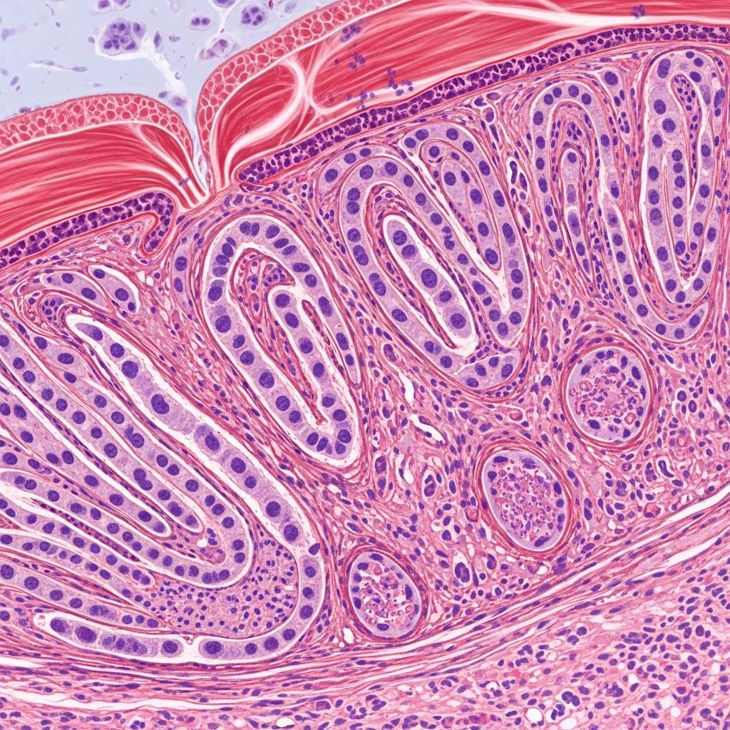
Spindle cell tumors represent a significant category of neoplastic growths that develop in canine patients, originating from the connective tissues found in or beneath the skin. These tumors compose a notable portion of dermatological malignancies in dogs, affecting animals across various breeds and age groups. Understanding the nature of these growths, their presentation, and available treatment modalities is essential for pet owners and veterinary professionals alike.
What Are Spindle Cell Tumors?
Spindle cell tumors in dogs are characterized by the proliferation of elongated, spindle-shaped or fusiform cells that arise from connective tissue origins. These neoplasms exhibit considerable morphological diversity and can originate from multiple tissue types, making their classification and diagnosis challenging even for experienced veterinary pathologists. The cells appear stretched and narrow when viewed under microscopic examination, which is the basis for their distinctive nomenclature.
These tumors fall under the broader category of soft tissue sarcomas (STS), which account for approximately 15 percent of all skin tumors encountered in canine medicine. The classification of spindle cell tumors encompasses various histological subtypes, including hemangiopericytomas, fibrosarcomas, myoepitheliomas, and peripheral nerve sheath tumors. Each subtype possesses unique biological characteristics and clinical behavior patterns that influence prognosis and treatment strategies.
Prevalence and Epidemiological Patterns
The incidence of spindle cell tumors varies across different populations and geographic regions. Research demonstrates that canine spindle cell mammary tumors represent approximately 1% of all canine mammary tumors, with regional variations noted—ranging from 0.83% in Milan to 1.13% in Vila Real. This relatively low prevalence underscores the specialized nature of these neoplasms within the broader context of canine oncology.
Certain canine populations show increased susceptibility to spindle cell tumor development. Large breed dogs, including Irish setters, Irish wolfhounds, golden retrievers, and Doberman pinschers, demonstrate elevated risk profiles for fibrosarcoma development. The average age of presentation occurs around 10 years, though juvenile cases have been documented in dogs under one year of age, often presenting with more aggressive biological behavior and unfavorable prognoses.
Gender representation in spindle cell tumor populations varies by tumor location and histological type. Fibrosarcomas affecting the nasal cavity and oral cavity demonstrate a predilection for male dogs, while other spindle cell tumor subtypes may show different gender distributions. Breed representation in affected populations includes mongrels and German Shepherds as predominant groups, though the diversity of breeds affected indicates that genetic predisposition is not absolute.
Classification and Histological Subtypes
Spindle cell tumors encompass multiple histological categories, each with distinct pathological characteristics and clinical implications:
- Malignant Myoepitheliomas: The most common subtype, comprising 64% of malignant spindle cell tumors, these originate from myoepithelial cells and demonstrate variable clinical behavior
- Hemangiopericytomas: Vascular-origin tumors showing branching staghorn vessel patterns and perivascular characteristics
- Fibrosarcomas: Connective tissue-derived tumors with fibroblastic differentiation, representing approximately 2% of malignant spindle cell populations
- Peripheral Nerve Sheath Tumors: Originating from nerve tissue, these tumors may demonstrate perivascular whirling patterns and comprise about 3% of malignant cases
- Undifferentiated Sarcomas: Tumors lacking clear histological lineage, representing 5% of malignant spindle cell neoplasms
- Benign Variants: Including angiomas and benign myoepitheliomas, these demonstrate less aggressive biological behavior
Clinical Presentation and Physical Characteristics
Spindle cell tumors typically manifest as soft or firm lumps developing beneath the canine skin. The tumors are usually locally invasive, meaning tumor cells extend into surrounding tissues, though some variants demonstrate limited invasive capacity. Most malignant spindle cell tumors present as solitary lesions in 57% of cases, though multiple tumor development can occur.
Tumor dimensions vary considerably, with average sizes of 6.42 ± 3.92 centimeters recorded in research populations, ranging from tumors as small as 1 centimeter to massive growths exceeding 20 centimeters. The majority of malignant spindle cell tumors exceed 5 centimeters at presentation, placing them in the T3 classification category. Tumors may appear well-demarcated or ill-defined, and texture varies from smooth to irregular surfaces.
Most affected dogs present with intact reproductive status (61% in studied populations), suggesting that hormonal factors may influence tumor development in certain histological subtypes, particularly mammary-origin spindle cell tumors. The tumors typically develop as non-painful masses, though large tumors may cause discomfort or functional impairment depending on anatomical location.
Diagnostic Methodologies and Pathological Evaluation
Accurate diagnosis of spindle cell tumors requires a multi-step approach integrating clinical assessment, histopathological examination, and immunohistochemical analysis. Initial diagnosis based solely on hematoxylin and eosin (HE)-stained tissue sections frequently differs from final diagnoses after immunohistochemical (IHC) evaluation—with discrepancies occurring in 75% of malignant cases. This substantial discordance rate highlights the critical importance of comprehensive diagnostic protocols.
Histopathological Analysis: Initial microscopic examination reveals spindle-shaped cells arranged in various growth patterns, including fascicular arrays, whorled configurations, or myxoid matrices. Pathologists assess cellular morphology, mitotic index (MI), and tissue architecture to determine histological grade, which directly correlates with biological aggressiveness.
Immunohistochemical Characterization: IHC analysis employs antibodies targeting multiple cellular markers to definitively classify tumor histotype and origin. Commonly utilized markers include pancytokeratin, cytokeratin 14, p63, calponin, vimentin, Ki-67, CD31, desmin, myosin, smooth muscle actin, glial fibrillary acidic protein, and S-100 protein. These markers help differentiate between myoepithelial, vascular, muscular, and neural-origin tumors.
Additional immunological markers provide diagnostic specificity for particular subtypes. CD34 immunoreactivity combined with STAT6 positivity specifically indicates solitary fibrous tumors. Laminin immunostaining aids in identifying schwannomas and distinguishing peripheral nerve sheath tumors from other spindle cell populations. VWF (von Willebrand factor) positivity characterizes endothelial cells within vascular-pattern tumors.
Metastatic Potential and Staging Considerations
Despite their frequently large size at presentation, malignant spindle cell tumors demonstrate relatively modest metastatic potential. Among studied populations with staging information, lymph node metastatic disease appeared in only 6% of cases, while distant metastatic spread was documented in just 3% of evaluated animals. These favorable metastatic patterns contrast with the tumors’ often substantial dimensions.
Tumor-related mortality rates remain remarkably low given the malignant histological classification, with approximately 8% of affected dogs dying from spindle cell tumor-related causes. In dogs with multiple mammary tumors, the malignant spindle cell tumor frequently represents the sole malignant neoplasm in 67% of cases, suggesting that its presence does not necessarily predict development of additional malignancies.
Histological grading according to canine soft tissue sarcoma schemes correlates with clinical outcomes and patient age. Higher sarcoma grades demonstrate significant associations with increased patient age and greater tumor dimensions. These grading systems evaluate cellular differentiation, mitotic activity, and necrotic components to predict biological behavior.
Associated Risk Factors and Etiological Considerations
Multiple factors contribute to spindle cell tumor development in canine patients. Injection site reactions have been increasingly implicated in tumor genesis, with growing evidence linking vaccination and therapeutic injections to subsequent tumor formation. This association parallels the well-established relationship between post-vaccination sarcomas and feline patients.
Breed-specific predispositions suggest genetic susceptibility factors in certain lineages. Large breed dogs demonstrate enhanced risk, though toy and medium-sized breeds occasionally develop these neoplasms. Age represents a significant risk factor, with peak incidence occurring in middle-aged to senior dogs, though juvenile presentations occur with greater aggressiveness.
Reproductive status may influence development of certain spindle cell tumor subtypes, particularly those with myoepithelial or mammary differentiation, given the predominance of intact females in affected populations. However, the mechanism by which estrogen or progesterone exposure might facilitate tumor development remains incompletely understood.
Therapeutic Approaches and Treatment Options
Surgical excision represents the primary therapeutic modality for spindle cell tumors, with complete removal offering the best opportunity for disease control. The completeness of surgical excision significantly influences recurrence probability, with incomplete margins substantially increasing recurrence likelihood. Wide surgical margins are typically recommended to minimize residual neoplastic cell populations.
The prognosis for dogs with spindle cell tumors varies depending on multiple factors including tumor grade, anatomical location, and surgical margins achieved. Grade I tumors (low-grade) generally demonstrate favorable long-term outcomes following complete surgical removal, while Grade II and III tumors (intermediate and high-grade) may warrant consideration of adjunctive therapies.
Adjunctive radiation therapy may benefit patients with high-grade tumors, incompletely excised lesions, or anatomically challenging locations where wide margins cannot be achieved without functional compromise. Chemotherapy agents have been explored in canine soft tissue sarcoma management, though evidence for spindle cell tumors specifically remains limited in veterinary literature.
Prognosis and Long-Term Outcomes
The prognosis for dogs with spindle cell tumors is generally more favorable than for many other canine malignancies, particularly when tumors are completely excised and demonstrate low to intermediate histological grades. The relatively low metastatic propensity and modest tumor-related mortality rates provide encouraging prospects for affected animals.
Dogs undergoing complete surgical removal frequently achieve prolonged survival times, with many never experiencing tumor recurrence or metastatic disease during their remaining lifespan. In research populations evaluating long-term outcomes, most tumor-related deaths occurred in dogs with higher-grade tumors, underscoring the importance of accurate histological classification and grade determination.
Monitoring and Surveillance Protocols
Following surgical treatment, regular physical examinations and monitoring for local recurrence remain essential. Owners should perform frequent palpation of surgical sites and report any new lumps or growths to their veterinarian. Radiographic or ultrasonic imaging may be warranted if clinical concerns arise regarding potential local recurrence or metastatic disease.
Dogs with completely excised low-grade spindle cell tumors may require minimal surveillance beyond routine physical examinations. Conversely, animals with high-grade lesions, incomplete excision, or documented metastatic disease warrant more intensive monitoring protocols and consideration of follow-up imaging studies.
Frequently Asked Questions
Are spindle cell tumors contagious or hereditary?
Spindle cell tumors are neither contagious nor directly hereditary, though genetic predispositions in certain breeds may increase susceptibility. Environmental factors and injection-site reactions appear to play important roles in tumor genesis.
Can spindle cell tumors be prevented?
While complete prevention is impossible, minimizing injection-site trauma, maintaining appropriate vaccination protocols, and regular health monitoring may reduce risk. Prompt removal of suspicious masses before they enlarge can improve treatment outcomes.
What is the difference between benign and malignant spindle cell tumors?
Benign tumors grow slowly and do not spread, while malignant tumors demonstrate cellular atypia and possess metastatic potential. Histopathological examination distinguishes between these categories.
How long can dogs live after spindle cell tumor diagnosis?
Many dogs with completely excised spindle cell tumors live normal lifespans. Prognosis depends on tumor grade, completeness of surgical removal, and individual patient factors.
Is chemotherapy necessary for all spindle cell tumors?
Not all spindle cell tumors require chemotherapy. Low-grade, completely excised tumors may not benefit from adjunctive therapy, while high-grade or incompletely removed tumors may warrant additional treatment consideration.
Conclusion: Moving Forward with Diagnosis and Treatment
Spindle cell tumors represent a diverse group of neoplasms requiring sophisticated diagnostic approaches to ensure accurate classification and appropriate treatment planning. The relatively favorable prognosis associated with these tumors, particularly when identified and treated promptly, provides hope for affected canine patients and their families. Collaboration between veterinary oncologists and surgical specialists ensures optimal outcomes, while continued research advances understanding of tumor biology and therapeutic options.
References
- Canine Spindle Cell Mammary Tumor: A Retrospective Study — SAGE Publishing/Journal of Comparative Pathology. 2019. https://journals.sagepub.com/doi/10.1177/0300985819829522
- Canine Spindle Cell Tumor Mimicking Human Classical Hemangiopericytoma — National Institutes of Health/PMC. 2018. https://pmc.ncbi.nlm.nih.gov/articles/PMC6261813/
- Fibrosarcomas and Spindle Cell Tumors in Dogs — VCA Animal Hospitals. https://vcahospitals.com/know-your-pet/fibrosarcoma-and-spindle-cell-tumors-in-dogs
- Soft Tissue Sarcomas in Dogs — Cornell University College of Veterinary Medicine/Riney Canine Health Center. https://www.vet.cornell.edu/departments-centers-and-institutes/riney-canine-health-center/canine-health-information/soft-tissue-sarcomas-dogs
- Canine Soft-Tissue Sarcomas — DVM360. https://www.dvm360.com/view/canine-soft-tissue-sarcomas
- Connective Tissue Tumors in Animals — Merck Veterinary Manual. https://www.merckvetmanual.com/integumentary-system/tumors-of-the-skin-and-soft-tissues/connective-tissue-tumors-in-animals



