

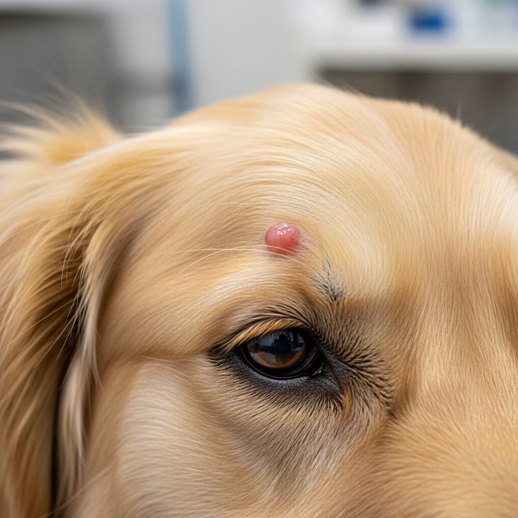
Understanding Basal Cell Tumors in Pets
Basal cell tumors represent one of the most common skin tumors found in both dogs and cats, particularly in middle-aged and older animals. These tumors develop when basal cells or other skin cells, such as those from sweat glands, hair follicles, or sebaceous glands, divide uncontrollably and cause an abnormal growth or mass on the skin. Understanding these tumors is essential for pet owners, as early detection and appropriate management can significantly improve outcomes for affected animals.
The good news is that most basal cell tumors are benign, meaning they are non-cancerous and pose limited health risks when managed properly. However, a small percentage can be malignant, requiring more aggressive treatment approaches. This distinction between benign and malignant forms makes proper diagnosis and veterinary evaluation crucial for any pet with a suspicious skin growth.
What Are Basal Cell Tumors?
Basal cell tumors are skin neoplasms that arise from the basal layer of the epidermis and related epithelial structures. With advances in immunohistochemical staining and veterinary pathology, our understanding of these tumors has evolved significantly. What was once classified under a single “basal cell tumor” umbrella term is now recognized as a heterogeneous group that includes several distinct tumor types, including trichoblastomas, basal cell carcinomas, and various sweat gland tumors.
The typical benign mass, which was previously called a basal cell tumor, is now recognized as a trichoblastoma. These tumors are characteristically well-circumscribed, firm masses that appear on the skin surface. In contrast, malignant basal cell carcinomas represent a separate entity with different biological behavior, growth patterns, and metastatic potential.
Who Is at Risk?
Basal cell tumors affect both dogs and cats, though the specific breed predispositions differ between species. Understanding which pets are at higher risk can help owners remain vigilant about monitoring their pets’ skin for any suspicious growths.
Canine Predisposition
In dogs, basal cell tumors are extremely common and most are benign. Canine basal cell tumors develop most commonly in middle-aged to older dogs, with several breeds showing increased susceptibility. Wirehaired Pointing Griffons, Kerry Blue Terriers, Wheaten Terriers, and Standard Poodles are among the breeds most predisposed to these tumors. In dogs, basal cell tumors are found most commonly on the head (especially the ears), the neck, and forelimbs.
Malignant basal cell carcinomas are less common in dogs than benign forms but can develop in older Standard Poodles and Cocker Spaniels. Saint Bernards, Scottish Terriers, and Norwegian Elkhounds face increased risk for the malignant variant, also known as basosquamous cell carcinoma in dogs.
Feline Predisposition
Basal cell tumors are particularly common in older cats. Domestic longhair, Himalayan, and Persian cats show the highest predisposition to these tumors. Cats can develop these tumors almost anywhere on their body, unlike dogs where certain locations are more common. Persians show an even higher tendency toward developing the malignant form, basal cell carcinoma, compared to other feline breeds.
Causes and Risk Factors
While veterinarians and researchers don’t fully understand why basal cell tumors develop, several factors have been identified that may increase a pet’s risk of developing these growths.
Environmental Exposure: Environmental factors, particularly sun exposure, may contribute to tumor development. Pets with lighter pigmentation and those spending significant time outdoors may face elevated risk. However, unlike squamous cell carcinomas, sun exposure is not considered the primary risk factor for basal cell tumors.
Genetic Predisposition: Genetics play a significant role in basal cell tumor development. The breed predispositions strongly suggest inherited factors that make certain dogs and cats more susceptible to these growths. Families with a history of skin tumors should monitor their pets more closely.
Age: Middle-aged and older pets are significantly more likely to develop basal cell tumors. Senior cats and dogs should be monitored regularly for any skin changes or new growths.
Other Factors: Scientists hypothesize that certain environmental exposures such as chemical toxins, specific viruses, or other unknown environmental factors may play a role in tumor development, though research in this area remains limited.
Clinical Presentation and Symptoms
Recognizing the typical appearance of basal cell tumors can help pet owners identify potential problems early. However, it’s important to note that visual appearance alone cannot definitively diagnose these tumors—veterinary examination and diagnostic testing are required.
Benign Basal Cell Tumors: These typically appear as firm, solitary, often hairless or ulcerated lumps on the skin. The lumps may stick out like stalks from the skin surface, creating a raised, nodular appearance. Benign tumors are usually slow-growing and may not cause visible symptoms beyond the physical growth itself.
Malignant Basal Cell Carcinomas: These malignant forms often appear as ulcers on the head, legs, or neck. Unlike benign tumors, malignant carcinomas are not usually raised above the skin surface. Instead, they tend to spread laterally through the skin, forming new ulcers as they progress. Some basal cell carcinomas may appear flattened or slightly raised, making them difficult to distinguish from benign forms without proper diagnostic testing.
Pet owners should watch for any lumps that grow rapidly, become ulcerated, or cause their pet to itch or scratch excessively at the affected area. Any changes in the appearance or behavior of a skin lesion warrant veterinary evaluation.
Diagnostic Procedures
Proper diagnosis is essential for determining whether a tumor is benign or malignant and for guiding appropriate treatment decisions. Veterinarians use several diagnostic methods to evaluate basal cell tumors.
Cytological Examination
Cytology is often the first diagnostic step and involves collecting cells from the tumor using a needle aspiration technique. The sample is then placed on a glass slide and examined under a microscope to identify the main cell type. In cytological diagnosis of basal cell origin tumors, cells are typically small, round to cuboidal, and arranged in tight clumps or ribbons.
However, it’s important to understand that cytologists may use the term “basal cell tumor” as a broad diagnosis covering multiple tumor types. This means cytology alone may not definitively distinguish between benign and malignant forms or identify the specific tumor type.
Histopathological Biopsy
When cytology cannot provide a definitive diagnosis, or when a tumor needs surgical removal, a biopsy offers more detailed information. Histopathology involves surgically removing a small piece of the tumor and sending it to a veterinary pathologist for microscopic evaluation. This procedure can accurately differentiate among trichoblastoma, basal cell carcinoma, sweat gland tumors, and other epithelial tumors.
A biopsy will definitively determine the tumor type and whether the tumor is benign or malignant. This information is crucial for developing an appropriate treatment plan and providing accurate prognostic information to pet owners.
Staging for Malignant Tumors
If histopathology reveals a basal cell carcinoma, complete staging is recommended to determine whether the cancer has spread. Staging procedures include locoregional lymph node aspiration, chest radiography, and abdominal ultrasonography. These tests help veterinarians assess the extent of disease and guide treatment planning.
Treatment Options
Treatment decisions depend on whether the tumor is benign or malignant, tumor size, location, and the pet’s overall health status. Different treatment approaches are available, ranging from conservative monitoring to aggressive multimodal therapy.
Benign Basal Cell Tumors
Surgical Excision: Most benign basal cell tumors diagnosed via cytology are easily cured with narrow surgical excision. Surgery involves removing the tumor along with a margin of normal surrounding tissue to ensure complete removal. For small, slow-growing tumors that aren’t bothering the pet, veterinarians may recommend a “wait and see” approach with periodic monitoring for growth or changes.
Monitoring: If a wait-and-see approach is chosen, both owner and veterinarian should periodically evaluate the mass for any growth or changes. Regular monitoring helps ensure that if the tumor begins to grow, becomes ulcerated, or causes the pet to itch or scratch excessively, treatment can be initiated promptly.
Malignant Basal Cell Carcinomas
Malignant basal cell carcinomas require more aggressive treatment and should be addressed as soon as possible to prevent spread and other negative effects. Malignant tumors in cats often display highly metastatic behavior, spreading to lymph nodes and occasionally to the lungs. Although reports are limited, veterinary oncologists have observed rare cases of metastasis in dogs.
Surgical Removal: Surgical removal is the primary treatment of choice for basal cell carcinomas. During surgery, the veterinarian removes a sufficient amount of normal skin around the tumor to ensure complete removal of all cancerous tissue. This approach may significantly reduce the risk of recurrence.
Cryosurgery: Specialty veterinary hospitals may offer cryosurgery, which uses low temperatures to freeze off the tumor. This technique can be effective for certain locations and tumor sizes.
Radiation Therapy: Radiation therapy may be recommended, particularly when surgical margins cannot be completely cleared or when the tumor location makes complete surgical removal difficult. Radiation helps eliminate remaining cancer cells after surgery.
Chemotherapy: When histology confirms a mass as malignant, chemotherapy may be of benefit. However, the malignant variant is rare enough that comprehensive studies evaluating chemotherapy efficacy are limited. Multimodal therapy combining surgery, radiation, and chemotherapy may achieve the best results for aggressive tumors.
Prognosis and Outcomes
The prognosis for basal cell tumors varies significantly based on whether the tumor is benign or malignant, the size at diagnosis, and the completeness of treatment.
Benign Tumors: The prognosis for benign basal cell tumors is generally excellent when caught early and managed properly. These tumors usually pose a limited risk to the pet’s well-being. Most pets enjoy a normal lifespan and quality of life following appropriate treatment.
Malignant Tumors: Malignant basal cell tumors (feline basal cell carcinoma) can also have a very good prognosis, particularly when caught early. These tumors spread to neighboring skin but seldom spread to other organs, which limits their systemic impact. Early detection and prompt appropriate treatment significantly improve the chance of full recovery. The earlier the tumor is identified and treatment initiated, the better the outcome.
Prevention and Monitoring
While not all basal cell tumors can be prevented, pet owners can take steps to minimize risk and ensure early detection. Regular skin examinations, particularly for pets in high-risk breeds or those over middle age, allow owners to identify new growths promptly. Limiting excessive sun exposure for outdoor pets may help reduce overall skin tumor risk. Additionally, maintaining a healthy lifestyle with proper nutrition and regular veterinary care supports overall skin health.
Frequently Asked Questions
Q: Are basal cell tumors contagious between pets?
A: No, basal cell tumors are not contagious. They develop from a pet’s own skin cells and cannot spread from one animal to another.
Q: How quickly do basal cell tumors grow?
A: Benign basal cell tumors are usually slow-growing, while malignant basal cell carcinomas may grow more rapidly. Some benign tumors may remain relatively stable for extended periods, while others may gradually enlarge over time.
Q: Can basal cell tumors return after surgical removal?
A: Recurrence is possible if the entire tumor is not completely removed during surgery. This is why veterinarians recommend removing adequate margins of normal tissue around the tumor. Histopathological confirmation of complete removal helps reduce recurrence risk.
Q: What should I do if I notice a new lump on my pet’s skin?
A: Schedule a veterinary appointment for evaluation. Your veterinarian can perform a physical examination and recommend appropriate diagnostic testing to determine whether the lump requires treatment.
Q: Are certain pet breeds at higher risk for malignant basal cell carcinomas?
A: Yes, certain breeds show increased predisposition. In dogs, Standard Poodles, Cocker Spaniels, Saint Bernards, Scottish Terriers, and Norwegian Elkhounds face higher risk. In cats, Persians are particularly predisposed to the malignant form.
Q: Is chemotherapy effective for basal cell carcinomas?
A: While chemotherapy may be of benefit when malignancy is confirmed, comprehensive efficacy studies are limited due to the rarity of malignant basal cell tumors. Your veterinary oncologist can discuss whether chemotherapy is appropriate for your pet’s specific situation.
References
- Cytology: Common Neoplastic Skin Lesions in Dogs & Cats — Today’s Veterinary Practice. 2024. https://todaysveterinarypractice.com/dermatology/common-neoplastic-skin-lesions-dogs-catscytologic-diagnosis-treatment-options/
- Basal Cell Tumors in Cats — PetCure Oncology. 2024. https://petcureoncology.com/basal-cell-tumors-in-cats/
- Tumors of the Skin in Dogs — MSD Veterinary Manual. 2024. https://www.msdvetmanual.com/dog-owners/skin-disorders-of-dogs/tumors-of-the-skin-in-dogs
- Basal Cell Tumors in Dogs — PetMD. 2024. https://www.petmd.com/dog/conditions/skin/basal-cell-tumors-dogs
- Tumors of the Skin in Cats — Merck Veterinary Manual. 2024. https://www.merckvetmanual.com/cat-owners/skin-disorders-of-cats/tumors-of-the-skin-in-cats
- Basal cell tumor or cutaneous basilar epithelial neoplasm — PubMed/National Center for Biotechnology Information. 2006. https://pubmed.ncbi.nlm.nih.gov/17123253/
- Basaloid epithelial tumors of dogs and cats — Veterinary Clinical Pathology. 2010. https://www.ovid.com/journals/vetcp/fulltext/10.1111/j.1939-165x.2010.00232_1.x



