
Reptile Veterinary Procedures: Essential Techniques For Vets
Comprehensive guide to essential clinical techniques for diagnosing and treating reptiles in veterinary practice.
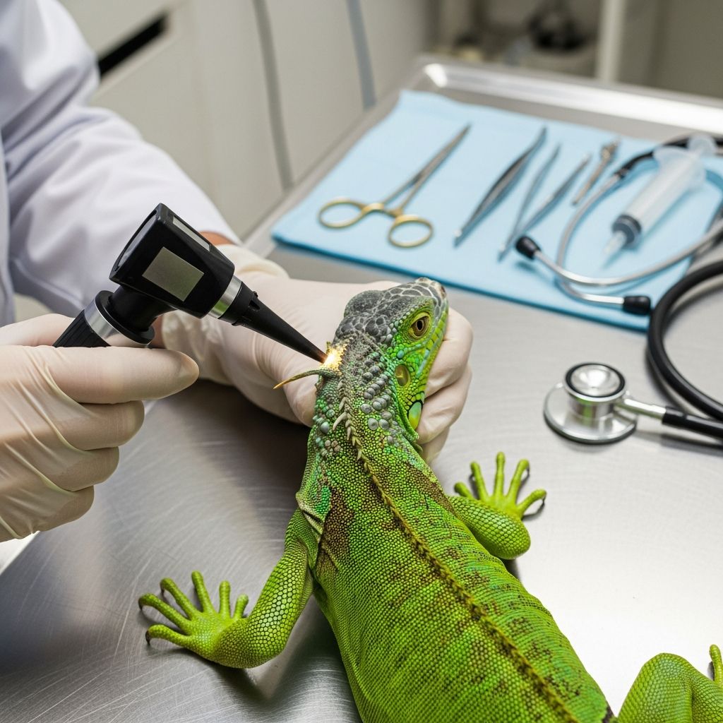
Reptiles, including turtles, tortoises, lizards, and snakes, require specialized veterinary approaches due to their unique physiology and behaviors. Effective clinical management begins with thorough assessments and progresses to advanced interventions when needed. This guide details key techniques used by veterinarians to maintain reptile health and address illnesses.
Initial Assessment and Observation Techniques
The foundation of reptile care lies in systematic observation before any hands-on contact. Start with a distant evaluation to note behavior, posture, and environmental interactions. Look for signs like lethargy, abnormal respiration, or substrate avoidance, which often indicate underlying issues. Fecal analysis during this phase can reveal parasites or digestive problems early on.
For chelonians, observe shell integrity and limb movement. Lizards may show tail abnormalities or limb swelling, while snakes require scrutiny of scale patterns and locomotion smoothness. Accurate body weight measurement is crucial, though challenging in large or stressed individuals. Hydration status assessment is vital, particularly post-hibernation or in debilitated patients.
Hands-On Physical Examinations
Transition to tactile exams after initial observations. Reptiles mask illness well, so detailed palpation and inspection are essential. Begin at the head and proceed caudally to minimize stress.
- Head Region: Check eyes for clarity, spectacles for retention (common in snakes and geckos), nares for discharge, and oral cavity for lesions, tongue health, and glottis position.
- Cardiovascular: Use Doppler ultrasonography on the prehumeral area or neck to assess heart rate and rhythm accurately, as traditional auscultation is unreliable due to scales or shell.
- Body Condition: Palpate dorsal musculature, coelom, and limbs. In chelonians, vertical restraint aids coelomic palpation for organ enlargement or eggs. Lizards need pelvic limb and tail checks; snakes focus on ventral scales.
- Vent and Tail: Inspect for prolapse, parasites, or trauma.
Small species may need only manual restraint, but larger ones often require sedation to prevent injury.
Restraint Methods for Safe Handling
Proper restraint prevents harm to both animal and handler. Physical methods include dorsal recumbency, dim lighting, or gentle ocular pressure to induce vasovagal responses for minor tasks like venipuncture. Chemical restraint is preferred for thorough exams or procedures.
| Reptile Type | Recommended Restraint | Key Considerations |
|---|---|---|
| Chelonians | Vertical positioning for coelom | Secure bridges to avoid scratches |
| Lizards | Tabletop with limb control | Sedation for active species |
| Snakes | Tube or manual pinning | Monitor for escape attempts |
Anesthesia and Sedation Protocols
Anesthesia is critical for invasive procedures. Inhalant agents are ideal, but injectables like propofol or alfaxalone via IV or intraosseous routes offer rapid induction with low toxicity. Premedication and preemptive analgesia improve outcomes. Monitor vital signs continuously, as reptiles have prolonged recovery times.
For minor interventions, sedation suffices. General anesthesia demands sterile technique and supportive care like temperature maintenance.
Diagnostic Imaging Modalities
Imaging enhances non-invasive diagnosis. Ultrasonography excels for soft tissues, guiding aspirations, and cardiac evaluation with color Doppler, though limited by air or bone. Transillumination with cold lights visualizes coelomic contents in small reptiles, detecting impactions.
Computed tomography (CT) provides superior detail for bone and soft tissues, often feasible with light sedation. MRI is useful for neurology but less common due to equipment needs.
Surgical Interventions in Reptiles
Surgery addresses trauma, reproductive issues, or research needs. Common procedures include coeliotomy, amputations, and shell repairs. Reptile skin is the primary strength layer, so everting sutures prevent dysecdysis post-incision. Suture removal occurs after shedding, around 6 weeks.
Feeding tube placement, like pharyngostomy, supports debilitated patients. Prep the neck, use a hemostat to access the pharynx, and secure the tube. For ophthalmic issues, spectacle wedge incisions or conjunctivoralostomy relieve blockages.
- Biopsy and device implantation for research.
- Ovariectomy or orchidectomy for population control.
- Fracture repairs using pins or external fixators.
Post-Procedure Recovery and Supportive Care
Recovery emphasizes thermoregulation, hydration, and nutrition. Provide heat gradients and monitor for complications like infection. Analgesia continues postoperatively, tailored to species.
Regular wellness exams promote early detection, as reptiles decline rapidly once symptomatic.
Common Challenges and Best Practices
Veterinarians face hurdles like poor venipuncture access and variable metabolism. Husbandry history informs differentials. Collaborate with specialists for complex cases.
Maintain sterility, use appropriate antibiotics, and document thoroughly.
Frequently Asked Questions (FAQs)
Do all reptiles need anesthesia for exams?
No, small or cooperative species may tolerate physical restraint, but sedation is advised for safety in most cases.
How do you assess hydration in reptiles?
Evaluate skin elasticity, oral mucus, and eye position. Weigh serially for trends.
What imaging is best for reptile GI issues?
Ultrasonography or CT; transillumination for small patients.
When is surgery necessary for reptiles?
For obstructions, reproductive diseases, trauma, or supportive care like tubes.
How long does reptile anesthesia recovery take?
Longer than mammals; hours to days, with monitoring.
References
- Performing Physical Examinations of Reptiles in General Practice — Today’s Veterinary Practice. 2023. https://todaysveterinarypractice.com/exotic-medicine/performing-physical-examinations-of-reptiles-in-general-practice/
- Laboratory Reptile Surgery: Principles and Techniques — PMC (NCBI). 2011-02-15. https://pmc.ncbi.nlm.nih.gov/articles/PMC3035398/
- Clinical Procedures for Reptiles — Merck Veterinary Manual. 2023. https://www.merckvetmanual.com/exotic-and-laboratory-animals/reptiles/clinical-procedures-for-reptiles
- Routine Health Care of Reptiles — MSD Veterinary Manual. 2023. https://www.msdvetmanual.com/all-other-pets/reptiles/routine-health-care-of-reptiles
- Essential Reptile Surgeries (Proceedings) — DVM360. 2019. https://www.dvm360.com/view/essential-reptile-surgeries-proceedings
- Guidelines on Reptile Anesthesia, Analgesia and Surgery — University of Michigan (UMCES). 2023. https://az.research.umich.edu/animalcare/guidelines/guidelines-reptile-anesthesia-analgesia-and-surgery/
Similar Articles







Read full bio of Sneha Tete