

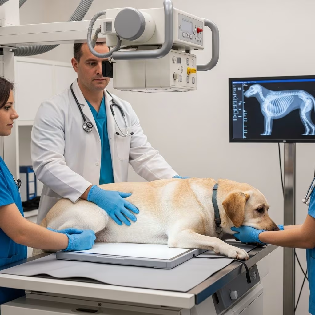
Understanding Radiographs for Dogs
Radiography, commonly known as x-ray imaging, is one of the most essential diagnostic tools available in modern veterinary medicine. When your veterinarian suspects an internal health issue in your dog, radiographs provide a non-invasive way to visualize bones, organs, and tissues beneath the skin. This comprehensive guide will help you understand what radiographs are, why they matter for your dog’s health, and what to expect during the procedure.
What Are Radiographs?
Radiographs are images created using electromagnetic radiation to penetrate tissues and create detailed pictures of your dog’s internal structures. During the radiographic process, x-rays pass through your dog’s body, and different tissues absorb varying amounts of radiation, which creates the image displayed on a digital screen or film. Bone appears white because it absorbs more radiation, soft tissues appear in shades of gray, and air-filled spaces like the lungs appear dark. Modern veterinary practices use digital radiographic systems, which offer superior image quality and significantly lower radiation exposure compared to traditional film-based systems.
Why Dogs Need Radiographs
Radiographs serve numerous diagnostic purposes in canine medicine. Your veterinarian may recommend radiographs to evaluate:
– Suspected fractures, bone diseases, or joint abnormalities- Internal organ function and structure- Abdominal distension or suspected foreign objects- Thoracic (chest) conditions like pneumonia or fluid accumulation- Spinal cord compression or intervertebral disc disease- Dental problems and tooth positioning- Suspected masses or tumors- Orthopedic conditions affecting limbs and joints
The ability to visualize internal structures without surgery makes radiographs an invaluable first-line diagnostic tool for determining the cause of your dog’s clinical signs.
Types of Radiographic Studies
Thoracic Radiographs
Thoracic radiographs evaluate the heart, lungs, and chest cavity. A complete thoracic radiographic study includes left lateral, right lateral, and ventrodorsal or dorsoventral views. Both right and left lateral recumbent radiographs are particularly important in dogs because they can reveal different pathologies depending on patient positioning. The thorax should be centered in the image, with the collimated field of view including the thoracic inlet cranially and extending to the caudal aspect of the pulmonary parenchyma and cranial abdomen caudally. This positioning ensures all relevant thoracic structures are captured for comprehensive evaluation.
Abdominal Radiographs
Abdominal radiographs are often used as a first-line screening test for possible intra-abdominal disease. A complete abdominal radiographic study should always include left and right lateral radiographs in addition to a ventrodorsal radiograph. Dorsoventral radiographs of the abdomen are hardly ever recommended because this position artifactually causes visceral crowding and secondary decreased serosal detail, thus rendering the radiograph essentially nondiagnostic.
Dogs are measured at the thickest part of their bodies, typically at the liver or cranial abdomen. For dogs measuring less than 15 cm, a reasonable starting technique would be 68 kVp and 8 mAs for a 400 film-screen analog film system, while digital plates require 72 kVp and 10 mAs. For dogs measuring 15 cm or greater, a grid should be used to reduce scatter radiation from degrading the final image quality.
Lateral radiographs of the abdomen should include the cranial aspect of the diaphragm and caudal thorax and extend to the coxal joints caudally. The margins of the cutaneous soft tissues should also be included. Radiographs of the abdomen are ideally made at peak expiration, if feasible.
Orthopedic Radiographs
Radiographs of any portion of the musculoskeletal system should always include at least 2 orthogonal radiographs. Intentionally oblique radiographs are commonly acquired to evaluate for instability, further characterize suspected lesions, or rule out specific differential diagnoses. It is imperative that straight, well-positioned radiographs of the musculoskeletal system are acquired; even subtle changes in obliquity can result in missed lesions, such as fracture planes. Patient positioning, collimation, appropriate positioning of a calibration marker, and knowledge of orthopedic landmarks are important factors in obtaining quality orthopedic radiographs.
Preparation for Radiographs
Proper preparation ensures your dog receives quality radiographs with minimal stress. Here are essential steps to prepare your dog for the procedure:
– Remove collars and metal items: Any metal objects, including collars, leashes, and ID tags, should be removed before radiographs as they can create artifacts on the image- Empty the bladder and bowels: For abdominal radiographs, your veterinarian may recommend a light enema or ask you to withhold food for several hours to reduce intestinal gas that might obscure organs- Maintain calm behavior: A relaxed dog produces better images; keep pre-procedure activities calm and reassuring- Follow fasting guidelines: If sedation will be used, your veterinarian will provide specific fasting instructions- Discuss medications: Inform your veterinarian about any medications your dog is currently taking- Bring relevant history: Provide information about your dog’s recent injuries, symptoms, or behavioral changes
The Radiographic Procedure
Understanding what happens during the radiographic procedure can help reduce anxiety for both you and your dog. The procedure typically follows these steps:
Positioning
Your veterinarian or veterinary technician will carefully position your dog to capture the necessary images. Proper positioning is critically important to maximize the diagnostic content of the x-ray examination. In many cases, improper positioning for radiographic examination can result in a misdiagnosis or inability to appreciate major lesions. For thoracic radiographs, both right and left lateral recumbent radiographs are recommended in dogs. The collimator light guides precise positioning, with specific horizontal and caudal borders depending on the anatomical area being examined.
Stabilization
Some dogs may require mild sedation or anesthesia to remain still during radiographic procedures, particularly for orthopedic or detailed anatomical studies. Your veterinarian will discuss sedation options if necessary. Proper restraint and positioning devices help minimize motion blur and ensure diagnostic quality images.
Radiation Exposure
Modern digital radiographic systems have wide exposure latitude, meaning mild changes in kilovoltage potential and milliampere-seconds tend to have minimal effect on the resultant images. This advancement allows veterinarians to optimize image quality while minimizing radiation exposure to your dog. Lead aprons protect areas not being radiographed, and multiple views are obtained to ensure comprehensive evaluation.
Image Interpretation
After radiographs are taken, your veterinarian interprets the images to identify abnormalities and formulate a diagnosis. Image viewing and interpretation should be performed on diagnostic monitors in a dark or dimly lit room. Imaging changes are often characterized by alterations in size, shape, location/position, opacity, and margination, which represent the basis of radiographic interpretation.
Knowledge of the history and signalment is absolutely necessary to achieve consistent and accurate interpretation of radiographic studies. Your veterinarian combines radiographic findings with clinical examination results and laboratory data to develop an accurate diagnosis and appropriate treatment plan. Board-certified radiologists and diagnostic imaging specialists are highly trained veterinarians who have spent considerable time beyond veterinary schooling to become experts in image interpretation. For complex cases, your veterinarian may consult with a board-certified veterinary radiologist through teleradiology services.
Special Radiographic Techniques
Contrast Radiography
Some diagnostic situations require contrast agents to improve visualization of specific organs or structures. Contrast radiography procedures include detailed explanations of indications, contraindications, and common pitfalls. These procedures may involve oral, intravenous, or rectal administration of contrast medium and are particularly useful for evaluating the gastrointestinal tract, urinary system, and vascular structures.
Dental Radiographs
Veterinary dental radiography uses specialized positioning techniques and smaller sensors or film sizes to capture detailed images of teeth, root structures, and surrounding bone. Full mouth radiographic series can often be completed in 15 minutes or less for large dogs and 6 minutes or less for small dogs and cats with proper positioning techniques and templates.
Stressed and Oblique Radiographs
Stressed, flexed, and oblique radiographs can be intentionally made to evaluate for instability, further characterize suspected lesions, or rule out specific differential diagnoses. Obliqued or stressed radiographs should be labeled with the obliqued direction and which area stress was applied to, respectively.
Digital Versus Film Radiography
The veterinary imaging field has largely transitioned to digital radiographic systems, which offer significant advantages over traditional film-based systems. Digital radiography provides immediate image availability, allowing for quick assessment and retakes if needed. The wide exposure latitude of digital systems means that images maintain diagnostic quality even with minor technique variations. Additionally, digital images can be enhanced, magnified, and easily stored in your dog’s medical records for future comparison.
Safety Considerations
Radiographs use ionizing radiation, but the radiation exposure from diagnostic radiography is considered safe when performed appropriately. Modern digital systems minimize radiation exposure significantly compared to older film-based systems. Your veterinary team follows strict safety protocols, including:
– Using the minimum radiation necessary to produce diagnostic images- Protecting non-target areas with lead shielding- Positioning dogs carefully to minimize repeat procedures- Using collimators to limit the radiation field to only necessary areas- Maintaining equipment regularly to ensure optimal function
Cost and Time Considerations
Radiographic procedures are relatively quick, typically taking 15 to 30 minutes depending on the number of views required and whether sedation is needed. The cost varies based on the number of radiographs, anatomical area being examined, and whether your veterinarian requires specialist interpretation. Many veterinary practices include radiographic services on-site, while others may refer complex cases to specialty imaging centers.
When Radiographs May Not Be Suitable
While radiographs are extremely valuable, some situations may require alternative imaging modalities. Ultrasound may be preferred for evaluating soft tissue organs, fluid accumulation, or pregnancy. Computed tomography (CT) and magnetic resonance imaging (MRI) provide more detailed cross-sectional images for complex neurological or orthopedic conditions. Your veterinarian will recommend the most appropriate imaging modality based on your dog’s specific condition and clinical presentation.
After Radiographic Examination
After radiographs are completed, your veterinarian will discuss the findings with you. An imaging report is typically comprised of 2 or 3 sections: a description of the radiographic findings, an interpretation or impression, and sometimes recommendations as to additional imaging or suggestions on how to make a final diagnosis. Depending on the findings and your dog’s clinical condition, your veterinarian may recommend follow-up appointments, additional diagnostic testing, or treatment options.
Establishing Technique Protocols
Establishing a technique chart to take radiographs makes it easy for the operator to arrive at a technique by simply correcting a standardized protocol for the size of the animal being examined and the anatomical area under consideration. This ensures that radiographs of the same anatomical region will have a consistent appearance from animal to animal, facilitating accurate comparisons over time and improving diagnostic accuracy.
Frequently Asked Questions (FAQs)
Q: Is it painful for dogs to have radiographs taken?
A: No, radiographs are completely painless. Dogs may feel mild discomfort only if they have an existing injury in the area being radiographed. The positioning required for proper imaging may cause some temporary discomfort for dogs with acute injuries, which is why sedation or anesthesia is sometimes recommended.
Q: How often can dogs safely have radiographs?
A: Dogs can have radiographs as needed for diagnostic purposes without risk of harm from radiation exposure at diagnostic levels. Modern digital systems use minimal radiation. Your veterinarian will recommend radiographs only when clinically necessary.
Q: Can radiographs detect cancer in dogs?
A: Radiographs can help identify some cancers, particularly those affecting bones or visible as masses within the chest or abdomen. However, ultrasound, CT, or biopsy may be needed for definitive diagnosis of certain cancers.
Q: Will my dog need to fast before radiographs?
A: Fasting requirements depend on the type of radiograph and whether sedation will be used. For abdominal radiographs without sedation, fasting may not be necessary, but your veterinarian will provide specific pre-procedure instructions.
Q: How long does it take to get radiograph results?
A: Digital radiographs are immediately available for viewing. Your veterinarian can often provide preliminary results on the same day. If specialist interpretation is needed, results may take 24-48 hours.
References



