

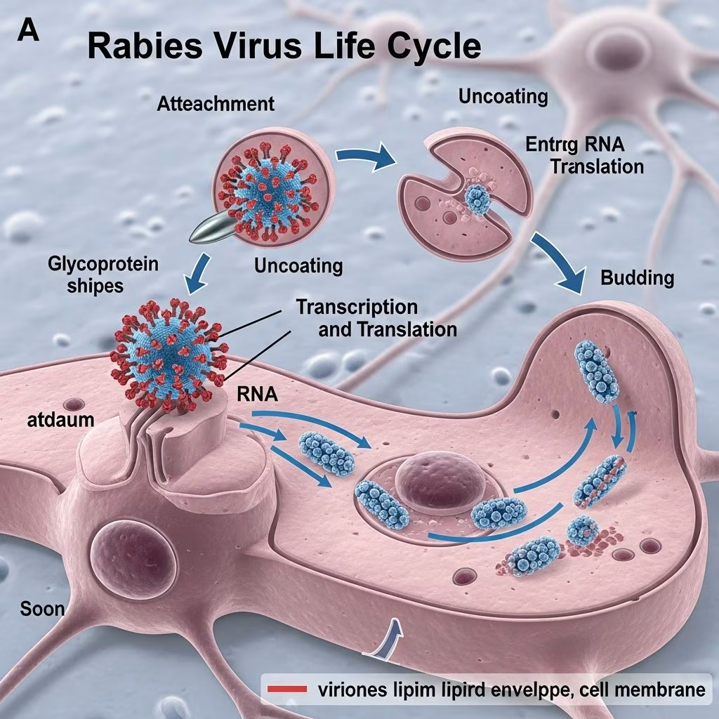
The rabies virus, a member of the Rhabdoviridae family, follows a precise sequence of events to infect hosts, replicate, and transmit. This negative-sense single-stranded RNA virus targets the nervous system, leading to almost certain death once symptoms appear. Understanding its life cycle is crucial for prevention strategies in veterinary and human medicine.
Viral Structure and Initial Host Interaction
Rabies virions exhibit a distinctive bullet-shaped morphology, measuring approximately 200 nm in length and 80 nm in width, enveloped by a host-derived lipid membrane. The envelope embeds the glycoprotein (G), which facilitates attachment to host cell receptors such as nicotinic acetylcholine receptors, neural cell adhesion molecule (NCAM), and p75 neurotrophin receptor. This flexibility in receptor binding allows infection across diverse cell types, particularly in muscle and nerve tissues.
Transmission typically occurs through bites from infected mammals like dogs, bats, raccoons, and foxes, introducing saliva containing high viral loads into wounds. The virus remains viable at the inoculation site—often muscle tissue—for days to months, evading immediate immune detection due to low replication rates initially.
- Glycoprotein G role: Mediates binding and fusion, critical for pathogenesis.
- Envelope composition: Host lipids studded with G spikes.
- Core components: Helical nucleocapsid with RNA genome encapsidated by nucleoprotein (N), plus phosphoprotein (P), matrix protein (M), and large polymerase (L).
Cellular Entry and Uncoating
Upon binding, the virus enters host cells via receptor-mediated endocytosis, specifically clathrin-coated pits, forming an endosome. The endosomal acidification (low pH around 5-6) triggers conformational changes in glycoprotein G, driving fusion between viral and endosomal membranes. This releases the ribonucleoprotein (RNP) complex—genomic RNA bound to N, P, and L—into the cytosol.
No nuclear involvement occurs; all processes unfold in the cytoplasm. The RNP serves as the template for subsequent transcription, as free RNA cannot be recognized by the viral polymerase.
| Entry Phase | Key Mechanism | Outcome |
|---|---|---|
| Receptor Binding | G protein interaction | Endocytosis initiation |
| Endosomal Fusion | pH-induced G change | RNP release to cytosol |
| Uncoating | Envelope shedding | Genome accessible for transcription |
Intracellular Transcription and Replication
Once in the cytosol, the viral polymerase complex (L with P cofactor) initiates primary transcription. It synthesizes five monocistronic mRNAs from the negative-sense genome, corresponding to N, P, M, G, and L genes. These mRNAs, capped and polyadenylated, are exported for translation on free ribosomes, producing viral proteins.
Matrix protein M regulates transcription levels and aids later assembly. After sufficient proteins accumulate, polymerase switches to replication mode, ignoring intergenic signals to produce full-length antigenome (positive-sense RNA). This antigenome, encapsidated by N, templates new negative-sense genomic RNAs, forming RNPs for progeny virions.
- Transcription: Produces mRNAs for protein synthesis.
- Replication: Yields antigenomes and genomes.
- Regulation: M protein balances transcription vs. replication; P acts as interferon antagonist.
This phase occurs efficiently in muscle cells initially, though replication is limited compared to neurons.
Neuronal Invasion and Centripetal Spread
From muscle, virions cross neuromuscular junctions into peripheral nerves. The virus hijacks dynein motors via P protein binding to DYNLL1, enabling fast retrograde axonal transport at 50-100 mm/day toward the central nervous system (CNS). This directional movement along microtubules is pivotal for pathogenesis, allowing evasion of humoral immunity.
In the CNS—dorsal root ganglia, spinal cord, brainstem, and cerebrum—robust replication ensues, causing neuronal dysfunction. Incubation periods vary (weeks to years) based on bite location (closer to CNS shortens time), viral dose, and host species. Highly innervated sites accelerate progression.
Table summarizing spread dynamics:
| Stage | Transport Direction | Speed | Key Protein |
|---|---|---|---|
| Peripheral to CNS | Retrograde | 50-100 mm/day | P-DYNLL1 |
| CNS Amplification | Intraneuronal | N/A | Polymerase complex |
Centrifugal Spread and Peripheral Dissemination
Late infection sees anterograde transport from CNS to periphery via kinesin motors, infecting organs like skin, cornea, and crucially, salivary glands. High salivary viral titers enable transmission through bites or licks on mucosa. This bidirectional spread ensures perpetuation.
In reservoirs like bats or carnivores, adaptations prolong survival: overwintering in hibernating bats or age-structured transmission in carnivores sustain cycles despite lethality.
Assembly, Budding, and Release
New virions assemble at plasma membranes. M protein condenses RNPs with G-embedded lipids; budding occurs without cytolysis, preserving host cell function. Released enveloped particles infect neighboring cells or exit via saliva.
- Assembly sites: Intracytoplasmic membranes or plasma membrane.
- Budding process: M-RNP matrix drives envelopment.
- Yield: Progeny ready for new infection cycles.
Host Factors Influencing the Cycle
Incubation variability (2 days to 5+ years) aids persistence. In bats, prolonged asymptomatic phases (>200 days) facilitate dispersal. Carnivore dynamics rely on population density and extinction-recolonization. P protein’s interferon antagonism suppresses immunity, enhancing spread.
Prevention Implications
Knowledge of the cycle underscores post-exposure prophylaxis efficacy: wound cleaning removes virus, immunoglobulins neutralize at sites, and vaccines prime immunity before CNS entry. Pre-exposure vaccination protects high-risk groups.
Frequently Asked Questions (FAQs)
What is the incubation period for rabies?
Typically 1-3 months, but ranges from days to years depending on bite site and host.
How does rabies virus travel to the brain?
Via retrograde axonal transport using host dynein motors.
Can rabies replicate outside the nervous system?
Yes, initially in muscle, then nerves and salivary glands.
Why is rabies nearly always fatal?
CNS invasion triggers irreversible encephalitis before symptoms.
How is rabies transmitted?
Primarily bites; saliva contact with mucosa or wounds.
References
- Rabies virus — Wikipedia. 2023-10-15. https://en.wikipedia.org/wiki/Rabies_virus
- Figure 3. Rabies virus life cycle — Uniformed Services University of the Health Sciences. 2020. https://digitalcollections.lrc.usuhs.edu/digital/api/collection/p16005coll10/id/108707/download
- Rabies Virus — Jefferson University Research. 2019-06-01. https://research.jefferson.edu/content/dam/academic/research/researcher-labs/schnell-lab/schnell_paper_5.pdf
- The spread and evolution of rabies virus — PMC (NCBI). 2019-11-27. https://pmc.ncbi.nlm.nih.gov/articles/PMC6899062/
- Rabies in Animals — Merck Veterinary Manual. 2023. https://www.merckvetmanual.com/nervous-system/rabies/rabies-in-animals
- Rabies — Centers for Disease Control and Prevention (CDC). 2024-07-01. https://www.cdc.gov/yellow-book/hcp/travel-associated-infections-diseases/rabies.html



