

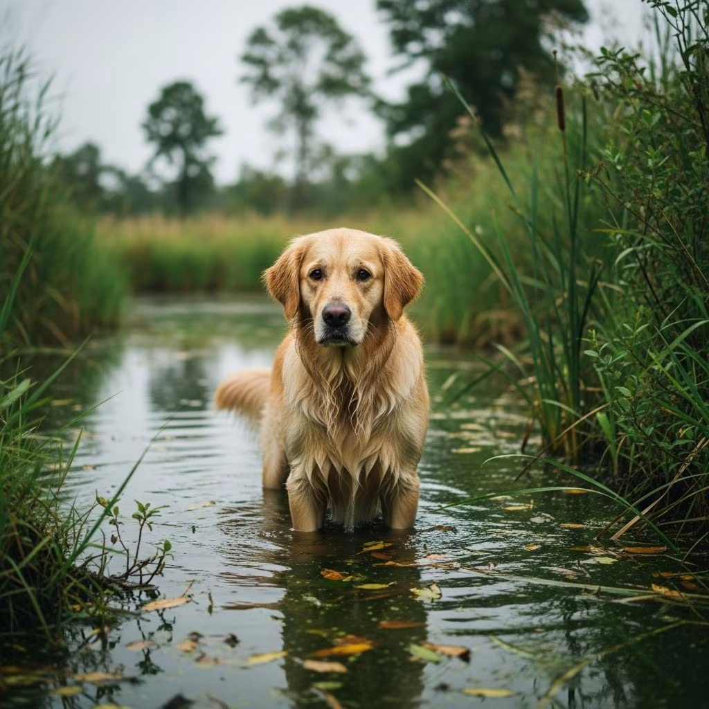
Pythiosis represents one of the most challenging and potentially life-threatening infections veterinarians encounter in canine medicine. This disease, caused by the aquatic organism Pythium insidiosum, manifests in dogs as a progressive and often fatal condition that demands immediate veterinary intervention. Unlike typical fungal infections, pythiosis behaves as both a fungal and parasitic pathogen, making it particularly difficult to treat with conventional antifungal medications alone. Understanding the nature of this disease, recognizing its symptoms, and knowing the available treatment options can significantly impact a dog’s chances of survival.
What Is Pythiosis and How Do Dogs Become Infected?
Pythium insidiosum is an aquatic oomycete—a water-dwelling organism that thrives in warm, moist environments such as lakes, ponds, swamps, and marshy areas. This pathogen is fundamentally different from true fungi, despite sharing similar characteristics that earned it the nickname “water mold.” The organism has historically been identified in tropical and subtropical regions, but emerging cases suggest its geographic range may be expanding.
Dogs typically contract pythiosis through direct contact with contaminated water or soil containing the organism’s zoospores—motile reproductive cells that seek out and penetrate the skin or mucosal membranes of the gastrointestinal tract. Most dogs possess a natural resistance to Pythium insidiosum, but those with compromised immune systems, genetic predispositions, or injuries that allow pathogen entry become vulnerable to infection. Young dogs, particularly those engaged in hunting or sporting activities that involve frequent water exposure, face elevated risk.
Clinical Manifestations: Two Primary Disease Forms
Pythiosis in dogs presents in two distinct forms: gastrointestinal and cutaneous. The gastrointestinal form affects the digestive tract, while the cutaneous form impacts the skin. Understanding the specific symptoms of each form is essential for early recognition and prompt treatment initiation.
Gastrointestinal Pythiosis
The gastrointestinal form represents the more common presentation of pythiosis in dogs. This variant causes progressive thickening of stomach and intestinal tissues, leading to partial or complete obstruction of the digestive tract. Affected dogs typically exhibit a constellation of symptoms that develop insidiously over weeks to months:
- Progressive weight loss and muscle wasting despite adequate food intake
- Chronic vomiting, often containing blood
- Diarrhea, frequently with visible blood in feces
- Difficulty defecating or straining during bowel movements
- Abdominal pain and visible discomfort
- Fever and general malaise
- Enlarged abdominal lymph nodes
- Decreased appetite and lethargy
A particularly challenging aspect of gastrointestinal pythiosis is that affected dogs often appear relatively healthy during the early stages of infection. By the time symptoms become apparent, the disease may have progressed significantly, making intervention more difficult.
Cutaneous Pythiosis
The cutaneous form manifests as severe skin lesions that fail to heal with standard wound care. Characteristic features include swollen, ulcerated nodules that drain persistently, creating open wounds resistant to treatment. The infection causes progressive tissue death, with affected areas developing a blackened appearance as necrosis advances. These lesions are typically invasive and may spread to surrounding tissues, requiring aggressive intervention to prevent systemic spread.
The Challenge of Early Diagnosis
Diagnosing pythiosis early presents considerable difficulty, as initial symptoms often appear non-specific. Weight loss combined with vomiting or diarrhea can indicate numerous gastrointestinal disorders, delaying specific diagnosis. Definitive diagnosis typically requires tissue sampling through biopsy and histopathological examination, where the characteristic tissue infiltrates and inflammatory patterns become visible under microscopic analysis.
Eosinophilic inflammation—a hallmark pathological finding in pythiosis—marks affected tissues with dense concentrations of specialized white blood cells. This intense inflammatory response directly contributes to the severe clinical signs and tissue damage observed in infected dogs. Serological testing for antibodies against Pythium insidiosum may provide supportive diagnostic information, though tissue examination remains the gold standard.
Surgical Intervention: The Primary Treatment Approach
Surgery represents the most effective initial treatment strategy for pythiosis. The goal of surgical intervention is complete removal of all infected tissue to eliminate the pathogenic load. The extent of surgical involvement depends on the disease location and severity.
For cutaneous pythiosis affecting limbs or the tail, surgical excision may require amputation of the affected appendage to achieve complete lesion removal. For gastrointestinal pythiosis, affected sections of the small or large intestine require surgical resection, followed by anastomosis (surgical reconnection of the remaining healthy bowel segments).
Following surgical removal, many veterinarians employ photoablation—a laser-based technique that destroys remaining fungal filaments in surrounding tissue that may escape surgical removal. This adjunctive approach aims to reduce recurrence risk by eliminating microscopic disease foci.
Medical Management and Antifungal Therapy
After surgical tissue removal, dogs receive prolonged antifungal medication protocols lasting from three to six months. The standard combination therapy employs itraconazole and terbinafine, ergosterol-targeting agents that inhibit the organism’s ability to incorporate essential sterols into its cell membranes. Some treatment protocols also incorporate amphotericin B administered intravenously for enhanced efficacy.
Unfortunately, antifungal therapy alone achieves successful disease resolution in only approximately 20 percent of treated dogs. This limited success rate reflects the fundamental nature of Pythium insidiosum—a pathogen that is not technically a fungus despite its fungal-like behavior and response to certain antifungal agents.
A 2020 case report examined the use of mefenoxam in combination with itraconazole and terbinafine. Results indicated that mefenoxam incorporation may improve outcomes, particularly for gastrointestinal pythiosis cases, though additional research is needed to establish definitive efficacy and optimal dosing protocols.
Emerging Treatment Protocols: Corticosteroids and Immunotherapy
Recent advances in pythiosis management have introduced novel treatment combinations that address both the inflammatory and immunological aspects of the disease. Research has demonstrated that low-dose prednisone or other corticosteroids, when combined with antifungal medications, may enhance treatment outcomes in dogs with non-resectable (surgically inaccessible) pythiosis.
Corticosteroids function in pythiosis treatment through multiple mechanisms. Eosinophils—the predominant inflammatory cells in pythiosis lesions—respond dramatically to corticosteroid treatment, exhibiting decreased adherence, inhibited chemotaxis, and apoptosis (programmed cell death). By reducing this intense eosinophilic inflammation, corticosteroids alleviate severe clinical signs while potentially enhancing the immune system’s ability to combat infection.
The most significant recent development in pythiosis management is the emergence of immunotherapy, specifically Pythium insidiosum antigen (PIA)—a therapeutic vaccine that fundamentally alters how a dog’s immune system responds to the pathogen. Unlike traditional preventive vaccines, PIA functions therapeutically to boost immunity in already-infected dogs. This immunological intervention represents the most promising treatment advancement, with complete disease resolution reported in dogs receiving this therapy.
Treatment Considerations for Non-Surgical Cases
In numerous cases, pythiosis lesions are so extensive and invasive that complete surgical removal is not feasible. In such situations, dogs receive extended courses of antifungal medications without surgical intervention. The most effective protocols combine early antifungals with low-dose prednisone and continuing immunotherapy, though success rates remain variable and prognosis remains guarded.
Nutritional Support and Supportive Care
Dogs with pythiosis typically experience severe weight loss and decreased appetite, necessitating specialized nutritional support throughout recovery. Veterinarians often recommend high-calorie, highly digestible food formulations that minimize gastrointestinal stress while maximizing nutrient absorption. Adequate nutrition proves essential for supporting immune function and facilitating tissue healing during the prolonged recovery period.
Dogs actively infected with pythiosis may require hospitalization with intravenous fluid support, particularly if they develop bowel obstruction or severe malnutrition. During hospitalization, careful monitoring of liver and kidney function becomes critical, as both antifungal medications and corticosteroids can cause hepatic and renal damage with prolonged use.
Prognosis and Long-Term Outcomes
The prognosis for canine pythiosis depends heavily on the timing of diagnosis and treatment initiation. Early detection when lesions remain localized offers substantially better outcomes than late-stage diagnosis with extensive systemic involvement. Dogs that undergo complete surgical excision followed by comprehensive medical management have improved survival chances compared to those with advanced disease at presentation.
Regular monitoring throughout treatment remains essential. Abdominal ultrasound imaging helps assess intestinal healing and detect recurrent disease in gastrointestinal pythiosis cases. Blood work monitoring ensures antifungal and corticosteroid therapies are not causing unacceptable organ damage. Serological titers may be tracked to assess immune response and treatment efficacy.
Prevention and Risk Reduction
While no preventive vaccine currently exists for pythiosis, risk reduction strategies focus on limiting water exposure in endemic regions. Dogs in geographic areas where Pythium insidiosum has been documented should have restricted access to stagnant water sources, particularly during warm months when organism activity peaks. Prompt treatment of skin injuries or wounds that may provide pathogenic entry points is also advisable.
Frequently Asked Questions About Canine Pythiosis
Is pythiosis contagious between dogs?
No, pythiosis is not contagious between dogs. The disease requires direct exposure to the aquatic organism Pythium insidiosum in contaminated water or soil. However, multiple dogs in the same household with exposure to the same water sources may become infected through environmental contamination rather than dog-to-dog transmission.
Can humans contract pythiosis from infected dogs?
Human infections with Pythium insidiosum are exceptionally rare and have not been documented through contact with infected dogs. Humans may theoretically contract the organism through direct environmental exposure to contaminated water, but this remains extraordinarily uncommon.
What is the survival rate for dogs with pythiosis?
Survival rates vary based on disease form, stage at diagnosis, and treatment approach. Dogs with cutaneous pythiosis undergoing complete surgical excision have improved survival compared to those with gastrointestinal disease. Overall, prognosis remains guarded, with early diagnosis and aggressive multimodal treatment offering the best chances for recovery.
How long does pythiosis treatment typically last?
Antifungal treatment protocols generally extend from three to six months following surgical intervention. Dogs receiving immunotherapy may require additional treatment courses extending beyond this timeframe. Complete resolution may take six months to over one year depending on disease extent and treatment response.
References
- Successful management of 3 dogs with colonic pythiosis using antifungal and corticosteroid combination therapy — National Center for Biotechnology Information (NCBI/PMC). 2019. https://pmc.ncbi.nlm.nih.gov/articles/PMC6524395/
- Water Mold Infection (Pythiosis) in Dogs — PetMD. Accessed 2026. https://www.petmd.com/dog/conditions/infectious-parasitic/c_multi_pythiosis
- Pythiosis in Dogs: Signs, Symptoms, and Treatment — American Kennel Club. Accessed 2026. https://www.akc.org/expert-advice/health/pythiosis-in-dogs/
- Pythiosis or Lagenidiosis (Oomycosis) in Dogs — VCA Animal Hospitals. Accessed 2026. https://vcahospitals.com/know-your-pet/pythiosis-in-dogs
- Pythiosis in Pets: Symptoms, Causes & Treatment — Miravista Veterinary Clinic. Accessed 2026. https://miravistavets.com/pet-owners/pythiosis/



