

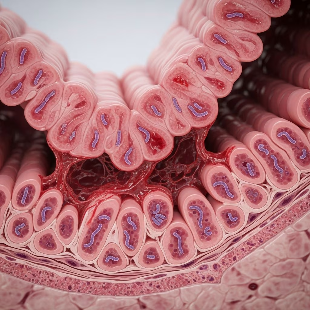
Porcine proliferative enteropathy (PPE), commonly referred to as ileitis, represents one of the most prevalent and economically significant intestinal disorders in modern pig farming. Triggered by the obligate intracellular bacterium Lawsonia intracellularis, this condition disrupts normal gut function, leading to a spectrum of outcomes from subtle growth setbacks to fatal hemorrhagic crises. Producers face challenges in managing this pathogen due to its ability to cause both overt disease and silent infections that erode profitability through reduced feed efficiency and uneven herd performance.
Understanding the Pathogen Behind PPE
Lawsonia intracellularis is a gram-negative, rod-shaped bacterium that thrives exclusively within the cytoplasm of intestinal epithelial cells in pigs. First successfully cultured in 1993 from infected porcine intestines, it fulfills Koch’s postulates through controlled challenge studies, confirming its role as the primary causative agent. This microbe targets rapidly dividing crypt cells in the ileum, jejunum, cecum, and colon, hijacking cellular machinery to propagate while halting normal cell maturation.
The bacterium’s lifecycle involves entry into enterocytes, where it replicates and spreads as host cells proliferate and migrate upward. This interference triggers hyperplasia of immature crypt cells, depletion of mature absorptive and secretory cells, and eventual mucosal thickening. Key signaling disruptions include upregulation of Notch-1 pathways and suppression of WNT/β-catenin signaling, which promote crypt cell overgrowth and impair goblet cell differentiation, reducing mucin production essential for gut barrier integrity.
Clinical Manifestations Across Age Groups
PPE exhibits diverse presentations influenced by pig age, immune status, bacterial dose, and concurrent stressors. Subclinical cases dominate, affecting grow-finish pigs with minimal diarrhea but notable impacts on average daily gain (ADG) and feed conversion ratios (FCR). Affected animals show variable body weights, apathy, and suboptimal market readiness.
- Chronic Form (Porcine Intestinal Adenomatosis – PIA): Typically strikes pigs aged 6-20 weeks post-weaning. Signs include pasty, gray-green diarrhea, weight loss, and poor condition without high mortality.
- Acute Hemorrhagic Form (Proliferative Hemorrhagic Enteropathy – PHE): Targets older growers or finishers (4-12 months), often naive replacements. Features sudden bloody diarrhea (tarry black to frank red), weakness, pallor, and mortality exceeding 50% in outbreaks.
- Complicated Forms: Necrotic enteritis involves secondary bacterial invasion leading to epithelial necrosis; regional ileitis features muscular hypertrophy and granulation from healing lesions.
In subclinical scenarios, herds experience ‘unevenness’ with some pigs thriving while others lag, collectively hiking production costs. Cold weather, high stocking densities, and gilt introductions heighten PHE risk.
Gut Pathology and Disease Progression
The hallmark of PPE is intestinal wall thickening due to unchecked crypt hyperplasia. Infected epithelia fail to differentiate, slashing absorptive surface area and protein uptake while spilling amino acids into the lumen—directly fueling diarrhea. Severe PHE cases reveal ileal and colonic mucosa engorged with blood, forming ‘ropes’ of clots upon necropsy.
| Clinical Form | Age Affected | Key Lesions | Mortality Risk |
|---|---|---|---|
| PIA (Chronic) | 6-20 weeks | Mucosal thickening, pasty feces | Low |
| PHE (Acute) | 4-12 months | Hemorrhagic ileum/colon, blood clots | High (>50%) |
| Subclinical | Grow-finish | Subtle hyperplasia, growth lag | None |
| NE/RI | Variable | Necrosis, muscular hypertrophy | Moderate |
Immune dynamics play a pivotal role: cellular immunity eventually clears most infections, but persistent carriers shed bacteria, perpetuating herd cycles. Co-factors like porcine circovirus type 2 (PCV2) or Salmonella amplify severity.
Diagnostic Approaches for Accurate Detection
Confirming PPE demands integrating history, clinical signs, and lab confirmation, as symptoms overlap with other enteropathies. Gross necropsy spots thickened, corrugated intestines; histopathology reveals silver-stainable intracellular rods amid hyperplastic crypts.
- PCR Testing: Quantitative PCR on feces detects bacterial DNA with high sensitivity, ideal for live herd screening and subclinical cases.
- Serology (ELISA): Identifies exposure via antibodies, useful for monitoring immunity but less specific for active infection.
- Histology/Culture: Gold standard postmortem, though culture is laborious due to the bacterium’s fastidious nature.
Challenges include asymptomatic shedders and variable shedding; routine surveillance via qPCR in high-risk groups (e.g., weaners) enables early intervention. Differential diagnoses encompass rotavirus, Clostridium, and coccidiosis.
Prevention and Control Strategies
Managing PPE hinges on biosecurity, vaccination, and targeted therapeutics. All-in-all-out production minimizes exposure; cleaning/disinfection targets resilient environmental forms.
- Vaccination: Injectable and oral vaccines prime mucosal immunity, slashing clinical disease in finishers. Administer per label to weaners or gilts.
- Antibiotics: Antimicrobials like tylosin or tiamulin during outbreaks curb bacterial replication; use judiciously to preserve efficacy.
- Management: Avoid mixing ages, monitor gilt health, and ensure nutritional balance to bolster immunity.
Eradication is feasible in clean herds via medicated early weaning or total depopulation-repopulation, though costly. Economic modeling underscores vaccination’s ROI through sustained ADG.
Economic Impact on Swine Operations
PPE drains profitability via treatment costs, mortality losses, and growth penalties. Subclinical infections alone can cut ADG by 20-30%, extending market time by days and inflating feed expenses. Outbreaks amplify culling and downtime. Proactive control averts multimillion-dollar hits in large operations.
FAQs on Porcine Proliferative Enteropathy
What causes porcine proliferative enteropathy?
Lawsonia intracellularis, an intracellular bacterium infecting intestinal crypts, is the sole cause.
How do you recognize PHE in pigs?
Look for acute bloody diarrhea, tarry feces, pallor, and sudden deaths in older pigs.
Is vaccination effective against ileitis?
Yes, both oral and parenteral vaccines significantly reduce clinical signs and improve growth.
Can subclinical PPE affect herd performance?
Absolutely; it causes uneven growth and poor feed efficiency without obvious diarrhea.
What is the best diagnostic test for live pigs?
Fecal qPCR for L. intracellularis DNA offers high sensitivity for early detection.
Swine producers must prioritize PPE surveillance to safeguard herd vitality. Integrating diagnostics, vaccination, and husbandry optimizes outcomes amid evolving production demands.
References
- Porcine proliferative enteropathy caused by Lawsonia intracellularis — Large Animal Review. 2023. https://www.largeanimalreview.com/index.php/lar/article/view/269
- Porcine proliferative enteropathy: overview of disease dynamics and host-bacteria interaction — PMC (PubMed Central). 2023-10-15. https://pmc.ncbi.nlm.nih.gov/articles/PMC12637199/
- Porcine Proliferative Enteropathy — Purdue Animal Disease Diagnostic Laboratory. 2004-06-01. https://www.addl.purdue.edu/newsletters/2004/summer/ppe.htm
- Ileitis – MSD Animal Health Swine — MSD Animal Health. 2024. https://www.msd-animal-health-swine.com/diseases-solutions/pigcare/intestipig/illeitis/
- Ileitis and Proliferative Enteropathies in Pigs — NADIS. 2023. https://www.nadis.org.uk/disease-a-z/pigs/ileitis-and-proliferative-enteropathies-in-pigs/



