

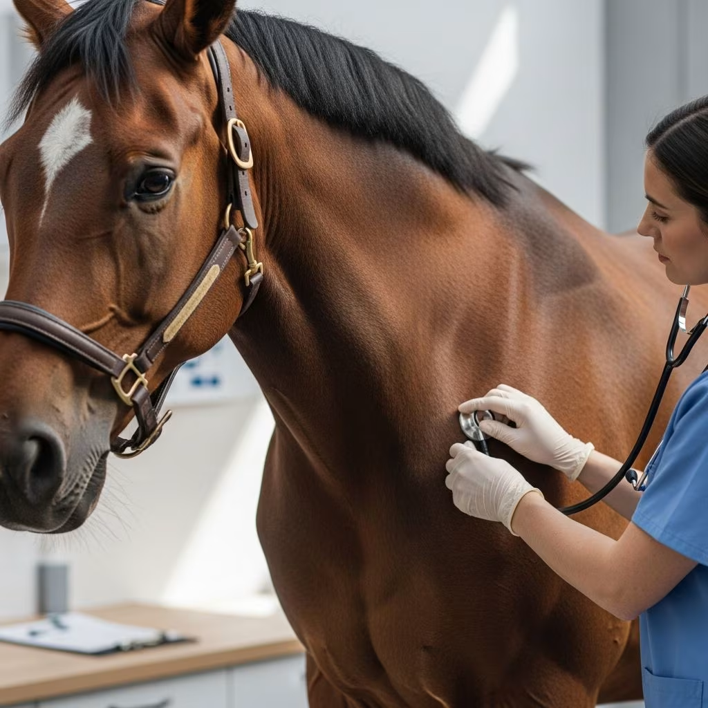
Pleuropneumonia, commonly known as **shipping fever**, represents a grave bacterial infection affecting both the lungs and the pleural cavity in horses. This condition arises when harmful microbes infiltrate the lower respiratory tract, leading to inflammation, fluid buildup, and potential organ dysfunction if untreated. Predominantly impacting performance horses under stress, it demands prompt veterinary intervention to avert fatal consequences.
Origins and Risk Factors Behind Equine Pleuropneumonia
The primary catalyst for pleuropneumonia is **long-distance transportation**, where horses endure prolonged trailer journeys with heads elevated and restricted. This setup impairs natural drainage of upper airway secretions, fostering bacterial migration into the lungs within 12-24 hours. Symptoms may emerge as swiftly as 24 hours or up to a week post-travel.
Beyond travel, contributing elements include prior viral infections weakening lung defenses, general anesthesia, intense exercise, and chest trauma. Young athletic horses below five years old, such as racers and sport competitors, face heightened vulnerability due to compromised clearance mechanisms.
- Transport stress: Elevated head position hinders mucus expulsion, enabling bacterial aspiration.
- Immune suppression: Viral illnesses or exhaustion reduce pulmonary resistance.
- Physical insults: Penetrating wounds introduce pathogens directly.
Aspiration of oropharyngeal bacteria, including anaerobes like Bacteroides spp., sparks bronchopneumonia that spreads to pleural surfaces, inciting exudative effusion and fibrin deposition.
Recognizing the Stages of Disease Progression
Pleuropneumonia evolves through distinct phases: peracute, acute, and chronic, each with escalating severity.
Peracute Phase
Initial nonspecific indicators include **fever**, inappetence, lethargy, and exercise reluctance. Horses appear dull without overt respiratory distress, mimicking mild viral ailments.
Acute Phase
As infection intensifies, pronounced signs emerge: productive cough, mucopurulent nasal discharge (sometimes fetid signaling anaerobes), rapid shallow breathing, and **pleurodynia**—sharp pleural pain manifesting as a braced stance, abducted elbows, grunting on chest percussion, and trunk rigidity. Sternal edema signals substantial effusion.
Chronic Phase
Persistent cases develop pleural loculations, lung consolidation, and abscesses, yielding ventral dullness on auscultation, radiating heart sounds, and systemic inflammation. Recovery prolongs, risking performance deficits from scarring.
| Stage | Key Symptoms | Duration |
|---|---|---|
| Peracute | Fever, lethargy, anorexia | 0-24 hours |
| Acute | Cough, pleurodynia, dyspnea, nasal discharge | 1-7 days |
| Chronic | Effusion loculations, abscesses, weight loss | Weeks to months |
Diagnostic Approaches for Accurate Identification
Diagnosis hinges on clinical evaluation, imaging, and fluid analysis to differentiate from heart failure, neoplasia, or trauma-induced effusions.
- Physical exam: Detects tachypnea, harsh lung sounds ventrally, pleural friction rubs, and pain on percussion.
- Ultrasonography: Reveals pleural fluid, comet-tail artifacts, lung consolidation, or abscesses—gold standard for pockets.
- Thoracocentesis: Aspirates septic effusion (pH <7.2, elevated lactate, bacteria on cytology, foul odor).
- Tracheobronchial aspiration (TBA): Samples lower airways for culture/sensitivity.
- Radiography: Supplementary for parenchymal changes, though challenging in adults.
Complete blood count often shows neutrophilia, hyperglobulinemia, and fibrinogen elevation, corroborating infection.
Comprehensive Treatment Strategies
Management targets infection eradication, effusion removal, pain control, and complication prevention, often spanning weeks with hospitalization.
Antimicrobial Therapy
Initiate broad-spectrum coverage presuming mixed aerobic/anaerobic flora: penicillin + aminoglycoside (gentamicin) + metronidazole. Tailor via culture results; duration 2-6 weeks IV then oral.
Chest Drainage and Lavage
Indwelling bilateral chest tubes facilitate continuous pleural flushing with heparinized saline or polyionic fluids to dissolve fibrin and clear debris. Monitor daily output; remove post-resolution.
Supportive Measures
- NSAIDs: Flunixin meglumine for pain, inflammation, endotoxemia.
- Polymyxin B: Counters endotoxin effects.
- Omeprazole: Prevents stress ulcers.
- Foot icing: Mitigates laminitis risk.
- Nebulization: Delivers antibiotics to airways.
Surgical Interventions
Refractory cases warrant **thoracotomy** to excise fibrin, drain abscesses, or break loculations. Video-assisted thoracoscopy aids minimally invasive access.
Potential Complications and Long-Term Outlook
Untreated or delayed therapy invites laminitis (sawhorse stance, hoof heat), thrombophlebitis, pneumonia recurrence, and restrictive lung disease from adhesions. Survival exceeds 80% with early aggressive care, though chronic scarring impairs athletics in 20-30%.
Prognosis improves with swift diagnosis; mild cases resolve in days, severe ones demand months. Post-recovery monitoring via ultrasound ensures clearance.
Preventive Measures for Horse Owners
Mitigate risks through:
- Minimizing transport duration; allow head-low positions for drainage.
- Vaccinating against respiratory viruses.
- Quarantining post-travel; monitor for fever.
- Avoiding overexertion in stressed horses.
Frequently Asked Questions (FAQs)
What triggers pleuropneumonia most frequently?
Long-haul travel with head elevation tops the list, promoting bacterial aspiration.
How do I spot pleurodynia in my horse?
Look for reluctance to bend, grunting on chest touch, and elbow abduction.
Is surgery always needed?
No, most respond to medical drainage and antibiotics; reserve thoracotomy for chronic fibrinous cases.
Can pleuropneumonia recur?
Yes, if incompletely treated or with resistant bacteria; follow-up imaging is crucial.
What’s the survival rate?
High (80-90%) with prompt care, lower in advanced stages.
References
- Equine Pleuropneumonia — Mississippi State University College of Veterinary Medicine. 2016-08-19. https://www.vetmed.msstate.edu/sites/www.vetmed.msstate.edu/files/presentations/8.19.16%20Equine%20Pleuropneumonia%20(Brittney%20Villagran).pdf
- Pleuropneumonia in Horses — Merck Veterinary Manual. 2023. https://www.merckvetmanual.com/respiratory-system/respiratory-diseases-of-horses/pleuropneumonia-in-horses
- Pleuropneumonia (Shipping Fever) in Horses — Vetster. 2024. https://vetster.com/en/conditions/horse/pleuropneumonia
- Pleuropneumonia (Shipping Fever) in Horses – Horse Owners — MSD Veterinary Manual. 2023. https://www.msdvetmanual.com/horse-owners/lung-and-airway-disorders-of-horses/pleuropneumonia-shipping-fever-in-horses
- Equine Pleuropneumonia — Baker McVeigh International. 2022. https://www.bakermcveigh.com/blog2/baker-mcveigh/equine-hospital/vet/blog-post-title-four-83m6s-z2wed-hpgx3
- Managing pneumonia and pleuropneumonia in horses (Proceedings) — dvm360. 2021. https://www.dvm360.com/view/managing-pneumonia-and-pleuropneumonia-horses-proceedings



