

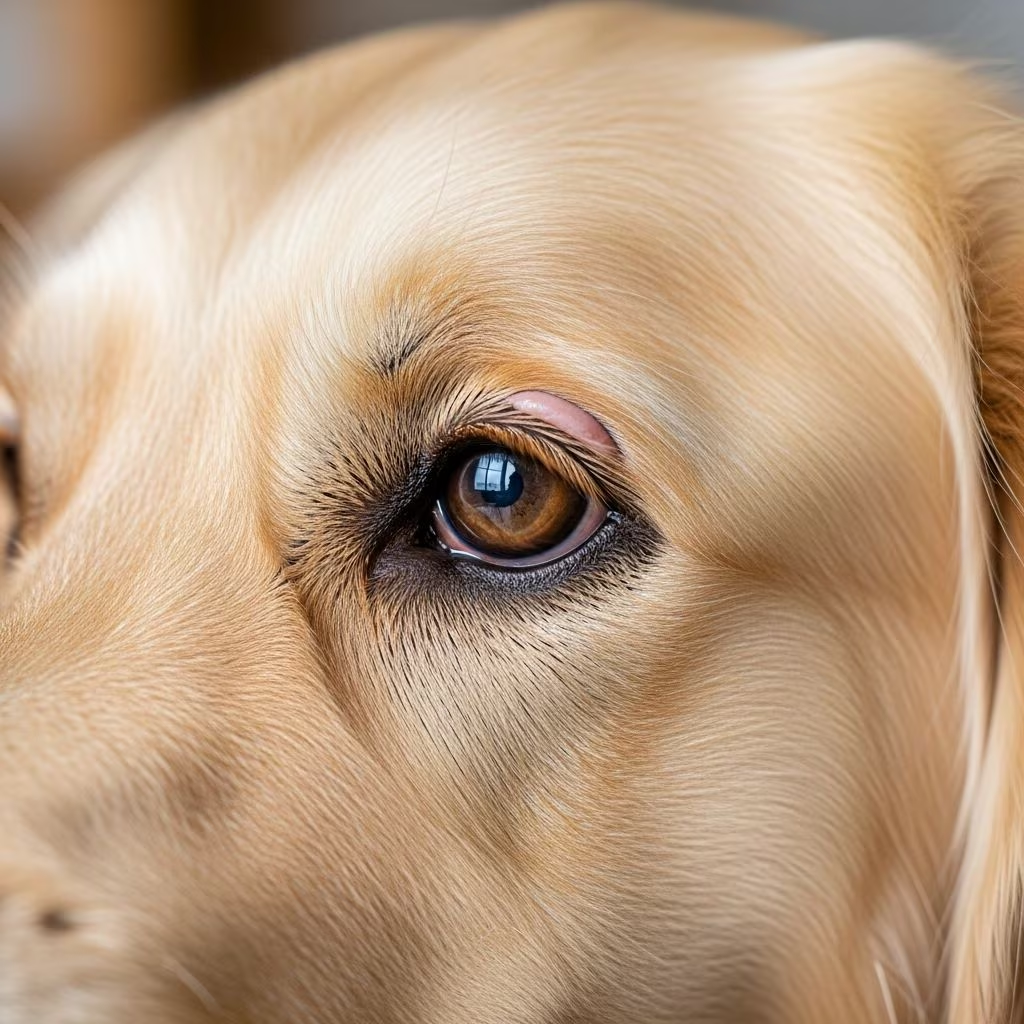
Most pet owners are familiar with the upper and lower eyelids that frame their dog’s or cat’s eyes, but few realize there’s a third eyelid hiding in plain sight. This remarkable anatomical feature, known scientifically as the nictitating membrane, plays a crucial role in maintaining eye health and protecting vision. Despite its importance, the third eyelid often goes unnoticed until something goes wrong. By understanding this hidden guardian of your pet’s eyes, you’ll be better equipped to recognize problems early and seek appropriate veterinary care.
The Hidden Architecture: What Is the Third Eyelid?
The third eyelid, also called the nictitating membrane or plica semilunaris, is a semi-transparent tissue structure located at the inner corner of your pet’s eye, tucked between the cornea and the lower eyelid. Unlike the upper and lower eyelids that move vertically, the third eyelid sweeps horizontally across the eyeball in a protective motion. In dogs and cats, this membrane typically remains largely hidden from view when the eye is healthy and relaxed, making it one of the eye’s most overlooked features.
The third eyelid consists of several distinct anatomical components working in harmony. A T-shaped cartilage provides structural support and rigidity, ensuring the membrane maintains its shape and function. Attached to the base of this cartilage is the gland of the third eyelid, a specialized tear-producing gland that contributes significantly to your pet’s tear film production. The entire structure is covered with conjunctiva, a mucous membrane that extends from the eyeball to the eyelids, creating a protective barrier.
Functional Roles: Why Your Pet Needs This Extra Eyelid
The third eyelid serves multiple critical functions that contribute to your pet’s ocular health and comfort. Understanding these roles helps explain why problems with this structure can lead to serious eye complications if left untreated.
Tear Film Production and Distribution
One of the most important functions of the third eyelid involves tear production. The gland of the third eyelid produces between 30 and 50 percent of the normal tear film in dogs, making it one of the most critical tear-producing structures in the eye. These tears are essential for maintaining a healthy eye surface, providing moisture, nutrients, and protective antimicrobial properties. The third eyelid doesn’t just produce tears; it also helps distribute them evenly across the eye surface as it sweeps across the eyeball, ensuring complete coverage and protection.
Corneal Protection
The cornea, the clear front surface of the eye, is one of the most sensitive and vulnerable parts of the visual system. The third eyelid acts as an additional protective layer, sweeping across the cornea during blinking and normal eye movement. This protective function becomes especially important during sleep or periods of reduced blinking, when the third eyelid helps maintain corneal moisture and shields against irritants.
Foreign Body Removal
As the third eyelid sweeps across the eye, it mechanically removes debris, dust, and other irritating particles from the eye surface. This cleansing action helps prevent foreign materials from scratching the cornea or causing inflammation. The space between the third eyelid and the eyeball serves as a common location where small foreign bodies may become lodged, which is why veterinarians routinely check this area during eye examinations.
Comparative Anatomy: How Different Species Use Their Third Eyelid
While dogs and cats both possess a third eyelid, the structure and function vary significantly across different animal species. Birds, for example, have a much more transparent third eyelid that they can control voluntarily, sweeping it across their eyes in a ventromedial direction. This voluntary control reflects the different visual demands of flight and predatory behavior. Interestingly, the avian third eyelid lacks an associated gland, relying instead on other tear-producing structures.
Many other mammals, including rodents and pigs, possess variations of the third eyelid as well. Some species even have additional specialized glands, such as the Harderian gland, located deeper within the orbit. These anatomical differences highlight how evolution has adapted the third eyelid to meet the specific needs of different species.
Common Conditions Affecting the Third Eyelid
When the third eyelid or its associated structures become diseased or injured, pet owners often notice visible changes. Several conditions can affect this important structure, requiring veterinary diagnosis and treatment.
Cherry Eye: The Most Recognizable Condition
The most commonly encountered third eyelid problem is a condition called cherry eye, which occurs when the gland of the third eyelid prolapses or slides out of its normal position. When this happens, owners notice a distinctive red, round mass appearing at the inner corner of the eye, resembling a cherry—hence the colloquial name. This condition can appear suddenly and is more common in young dogs of certain breeds, though cats can also develop it.
Cherry eye occurs when the tissues that normally hold the gland in place become weakened or fail to develop properly. The displaced gland becomes visible and often swells, creating the characteristic cherry-like appearance. While the sight of cherry eye can alarm pet owners, the condition itself is treatable. Modern veterinary medicine recommends repositioning the prolapsed gland rather than removing it entirely. This approach is preferable because removing the gland permanently increases the risk of dry eye disease, requiring lifelong medical management.
Cartilage Eversion and Scrolled Cartilage
Another condition affecting the third eyelid is cartilage eversion or scrolling, which occurs when the T-shaped cartilage becomes bent or twisted abnormally. This condition typically affects young giant breed dogs during their rapid growth phase and results in the third eyelid appearing as a pink or reddish mass in the corner of the eye. While scrolled cartilage may resemble cherry eye in appearance, the underlying cause is completely different, and veterinarians may need to perform examination under anesthesia to distinguish between these conditions and recommend appropriate treatment.
Amelanotic Leading Edge
Some individuals naturally have reduced or absent pigmentation on the free edge of the third eyelid, revealing normal, well-vascularized conjunctiva beneath. This condition, called an amelanotic leading edge, is not abnormal and requires no surgical correction. However, owners or breeders sometimes mistake this appearance for inflammation or disease. The unpigmented area becomes more noticeable when the eye is actually inflamed for other reasons, as the conjunctival blood vessels become more prominent and visible without the masking effect of pigment.
Recognizing When Something Is Wrong
While the third eyelid typically remains inconspicuous in healthy eyes, several signs indicate that veterinary attention is needed. If you notice the third eyelid becoming more visible than usual, appearing swollen or inflamed, or if you see a red mass at the inner corner of the eye, these changes warrant a veterinary examination. Excessive tearing, eye discharge, squinting, or apparent discomfort around the eye area may also indicate third eyelid problems or other eye conditions.
Additionally, conditions affecting the eyeball itself can cause secondary third eyelid protrusion. For example, glaucoma, a condition involving elevated intraocular pressure, can cause the eye to swell, making the third eyelid more prominent. Similarly, any condition causing the eyeball to retract slightly backward into the orbit may make the third eyelid appear more visible.
Consequences of Third Eyelid Removal
Historically, veterinarians sometimes recommended removing the third eyelid when it became problematic, but this approach is no longer recommended. The consequences of third eyelid removal can be significant and long-lasting. Removal of the gland associated with the third eyelid substantially increases the risk of developing dry eye disease, also called keratoconjunctivitis sicca. Without adequate tear production from this important gland, pets require lifelong medical management with artificial tears and other therapeutic agents to maintain corneal health and prevent painful complications.
Examination and Diagnosis
Veterinarians routinely examine the third eyelid during comprehensive eye examinations. To visualize the bulbar surface of the third eyelid and the space behind it, the veterinarian may gently manipulate the eyelid, reflecting it to examine the area thoroughly. This examination allows detection of foreign bodies, inflammation, or other abnormalities. In some cases, additional diagnostic techniques such as examination under anesthesia may be necessary to fully evaluate the structure and determine the nature of any problem.
Prevention and Maintenance
While some third eyelid conditions are hereditary or developmental and cannot be prevented, maintaining overall eye health helps prevent secondary problems. Regular eye examinations allow early detection of any issues. Keeping your pet’s face clean and free from irritants reduces the risk of conjunctival inflammation that could affect the third eyelid. If your pet has any breed predisposition to third eyelid problems, discussing preventive measures and early signs with your veterinarian is valuable.
Frequently Asked Questions About the Third Eyelid
Is it normal to see my pet’s third eyelid?
In healthy eyes, the third eyelid should remain mostly or completely hidden. If you notice it becoming visible, this typically indicates an underlying problem requiring veterinary evaluation. However, some unpigmented third eyelids may be more noticeable than others without being abnormal.
Can the third eyelid be surgically repaired if it’s damaged?
Yes, many third eyelid conditions can be surgically addressed. Prolapsed glands are typically repositioned rather than removed. Cartilage problems may require surgical correction depending on the severity and the pet’s comfort level.
Will my pet go blind if the third eyelid is removed?
While removal won’t cause immediate blindness, the loss of the tear-producing gland increases dry eye risk, which can lead to corneal damage and vision problems if not properly managed. This is why preserving the third eyelid is preferred whenever possible.
How do I know if my pet has an infection of the third eyelid?
Signs of infection include swelling, redness, discharge, and increased tearing. Your pet may also squint or show signs of eye discomfort. Professional veterinary examination is necessary to diagnose and treat infections properly.
Conclusion: Appreciating a Remarkable Structure
The third eyelid represents a remarkable evolutionary adaptation that contributes significantly to your pet’s eye health and comfort. From tear production to corneal protection and debris removal, this hidden structure performs essential functions that we often take for granted. By understanding its anatomy and recognizing signs of problems, pet owners can help ensure their companions maintain healthy vision throughout their lives. Should you notice any changes to your pet’s eyes or visible third eyelid, consulting with your veterinarian promptly ensures appropriate diagnosis and treatment, preserving this important structure for years of continued protection and comfort.
References
- Third Eyelid — Veterian Key. Accessed February 27, 2026. https://veteriankey.com/third-eyelid-2/
- Tales Of The Third Eyelid — South Texas Veterinary Ophthalmology. Accessed February 27, 2026. https://stvopets.com/articles/tales-of-the-third-eyelid/
- Nictitating Membrane — Wikipedia. Accessed February 27, 2026. https://en.wikipedia.org/wiki/Nictitating_membrane
- Conditions of the Third Eyelid Factsheet — Vet Specialists UK. Accessed February 27, 2026. https://www.vetspecialists.co.uk/fact-sheets-post/conditions-of-the-third-eyelid/
- The Anatomy and Function of a Cat’s Third Eyelid — All About Vision. Accessed February 27, 2026. https://www.allaboutvision.com/eye-care/pets-animals/cat-third-eyelid/



