

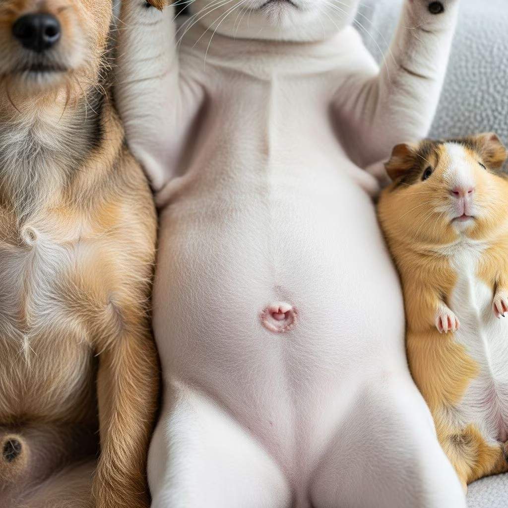
When you run your hands over your beloved dog or cat, you might occasionally wonder about the small marking on their abdomen—the belly button. While humans have prominent navels that are often discussed and even displayed, pet belly buttons remain a mystery to many owners. Understanding these tiny anatomical features can provide insight into your pet’s health and development, and in some cases, may reveal important medical considerations that require professional attention.
The Anatomical Foundation: What is a Belly Button?
A belly button, known scientifically as the umbilicus, represents a crucial connection during fetal development. Before birth, the umbilicus serves as an attachment point where blood vessels from the placenta connect to the developing fetus. This remarkable biological structure functions as an in utero life support system, delivering essential nutrients and oxygen while removing metabolic waste products. Without this connection, prenatal survival would be impossible.
The process of belly button formation differs significantly between humans and animals. In humans, medical professionals carefully manage the umbilical cord during delivery, typically placing clamps on the umbilical vessels before separating the placenta from the infant. These clamps remain in place until they naturally fall off several days after birth, leaving behind the characteristic navel we recognize in adults.
For pets, the situation is quite different. Mother dogs and cats instinctively chew through the umbilical cord after delivery, and the veterinarian’s role is typically absent during the birth process. This natural behavior results in a belly button that appears fundamentally different from human navels. In adult dogs and cats, the belly button is typically flat, without hair, and often resembles a small scar. Despite being covered in fur, pet belly buttons are relatively easy to locate, appearing as a cowlick—a small tuft of hair growing in an unusual direction on the abdomen.
Distinguishing Between Two Types: Formation and Appearance
Pet belly buttons come in two primary variants: innies and outies. An innie belly button is the more common type, where the navel appears flat or slightly indented into the abdominal wall. This configuration is seen in approximately 90% of humans and remains the predominant type in animals as well.
An outie belly button, conversely, protrudes slightly outward from the abdominal surface, creating a visible bump or extension. In humans, only about 10% of the population has an outie belly button. This rarity in humans carries through to animals, where outies are equally uncommon.
The determination of whether an individual—human or animal—develops an innie or outie is not something that can be controlled or predicted with certainty. Research involving human obstetrical professionals reveals an interesting fact: medical practitioners have no influence over whether a baby develops an innie or outie configuration. The outcome appears to be a natural consequence of how the body heals after the umbilical cord is severed and the umbilical stump falls away. This healing process involves factors that remain largely beyond medical intervention, suggesting that genetics and individual biological variation play determining roles.
The Rare Complication: Umbilical Hernias in Pets
While most pet belly buttons are simply anatomical variations without medical significance, some pets develop a condition called an umbilical hernia. Understanding this condition is crucial for pet owners, as it may require veterinary intervention.
An umbilical hernia occurs when the abdominal wall muscles fail to close completely around the umbilical cord during the developmental process, either in utero or shortly after birth. This muscular defect creates a small opening in the body wall near the umbilicus. When such a gap exists, tissue or even abdominal organs can potentially protrude through this opening, resulting in the characteristic outie appearance.
The severity of umbilical hernias varies considerably. In mild cases, the hernia causes no medical problems whatsoever and may go unnoticed by pet owners. The protruding tissue remains stable, and the pet experiences no discomfort or functional impairment. However, in more severe cases, abdominal organs can protrude through the hernia opening, which may necessitate surgical intervention to prevent serious complications.
When veterinarians identify umbilical hernias during physical examinations, the decision to repair surgically depends on several factors, including the hernia’s size, the pet’s age and overall health, and whether complications have developed. Many hernias that pose no current medical risk are repaired during routine surgical procedures such as spaying or neutering, taking advantage of the surgical access already required for these procedures.
Identifying Your Pet’s Belly Button Characteristics
Pet owners often find it challenging to locate their cat or dog’s belly button, particularly in animals with thick fur coats. Learning to identify and examine this small feature can help you become more familiar with your pet’s normal anatomy.
- Location: Pet belly buttons are located on the abdomen, typically positioned just below the ribcage, directly in the center of the belly area.
- Appearance in furred animals: Look for a small cowlick or tuft of hair that stands apart from the surrounding fur pattern. This distinctive hair growth pattern often marks the belly button’s location.
- Texture and color: When you locate the belly button, it typically appears as a small, flat scar-like marking. The skin may be pigmented differently from surrounding tissue, sometimes appearing lighter or forming a small line.
- Size reference: Pet belly buttons are significantly smaller and less prominent than human navels, often requiring deliberate searching to identify.
Gentle palpation—careful feeling of the area with your fingertips—can help you confirm you’ve found the correct location. Never force your pet to remain still if they’re uncomfortable, as the belly is often a sensitive area that some animals prefer not to have touched.
When Should You Be Concerned?
Most pet belly buttons require no special attention or care. However, certain signs warrant veterinary evaluation:
- Significant protrusion: If your pet’s belly button appears as a noticeable bump that seems larger than normal, professional assessment is advisable.
- Discharge or odor: Any unusual drainage, foul odor, or signs of infection around the umbilical area should be reported to your veterinarian.
- Pain or sensitivity: If your pet shows pain when the belly button area is touched or examined, this may indicate inflammation or infection.
- Changes over time: If the appearance of your pet’s belly button changes significantly, grows larger, or becomes inflamed, seek veterinary guidance.
- Behavioral changes: Excessive licking or biting at the belly button area, or signs of discomfort in that region, warrant professional evaluation.
Surgical Considerations and Recovery
When umbilical hernias require surgical repair, the procedure is typically straightforward. Veterinarians often repair these hernias during routine spay or neuter surgeries, as the abdominal incision required for these procedures provides convenient access to the umbilical region. The hernia repair involves closing the muscular defect and ensuring proper wound closure.
Recovery from umbilical hernia repair follows standard post-operative protocols. Pets require rest and restricted activity during the healing period, typically seven to fourteen days. Owners must prevent licking or chewing at the incision site, monitor for signs of infection, and follow all veterinary instructions regarding activity restrictions and wound care.
The Broader Context: Belly Button Diversity
The study of belly buttons extends beyond simple anatomy. Human research initiatives such as the Belly Button Biodiversity Project investigate the microbial communities that inhabit human navels, revealing fascinating insights about bacterial diversity and colonization patterns. While comparable research in veterinary medicine remains limited, these investigations highlight that belly buttons represent more than simple anatomical remnants—they are living, dynamic features worthy of scientific attention.
Frequently Asked Questions
Do all cats and dogs have belly buttons?
Yes, all cats and dogs have belly buttons, just like all mammals. However, they are typically much smaller and less obvious than human navels, and they are often hidden beneath fur.
Is it normal for a pet’s belly button to look different from mine?
Absolutely. Pet belly buttons are naturally different from human ones. They appear as flat scars rather than the prominent innies or outies seen in humans. This difference is completely normal and expected.
Can pregnancy affect a pet’s belly button appearance?
While pregnant female pets may experience some abdominal changes, the belly button itself typically does not show the dramatic changes sometimes seen in pregnant humans. Pregnant women may temporarily develop an outie appearance, but this is less common in animals.
Should I clean my pet’s belly button?
Pet belly buttons, unlike human innies, do not typically accumulate lint or require regular cleaning. The flat, scarred nature of pet navels means they do not have the infolded structure that can trap debris.
Is an outie belly button on my pet a sign of a serious problem?
Not necessarily. While an outie may indicate an umbilical hernia, many hernias cause no medical problems. However, any noticeable protrusion should be evaluated by a veterinarian to determine if treatment is necessary.
Conclusion: Appreciating Your Pet’s Unique Features
Your pet’s belly button, though small and often overlooked, represents a direct connection to their prenatal development and early life. Whether your dog or cat has an innie or the rarer outie configuration, this anatomical feature tells the story of their growth from fetus to newborn to adult companion. Understanding belly button basics empowers you to monitor your pet’s health effectively and recognize when veterinary consultation might be warranted. Most pets’ belly buttons require no special attention beyond the general awareness that comes from knowing your animal’s normal anatomy. However, remaining observant about changes or unusual characteristics ensures you can respond promptly should any health concerns arise. The next time you catch a glimpse of your pet’s belly button, you’ll appreciate not just the curiosity of this small feature, but also its significance in your companion’s biological story.
References
- The Difference Between an Outie and Innie – English Tutor Nick P — Tutor Nick P. June 12, 2025. https://www.youtube.com/watch?v=NArXpPAh0mQ
- Innie vs. Outie: Understanding Belly Button Types — Oreate AI Blog. Accessed February 2026. https://www.oreateai.com/blog/innie-vs-outie-understanding-belly-button-types/
- Does Your Pet Have an Innie or an Outie? — Animal Medical Center of New York. January 16, 2012. https://www.amcny.org/blog/2012/01/16/does-your-pet-have-an-innie-or-an-outie/
- Do Cats Have Belly Buttons? — PetMD. Accessed February 2026. https://www.petmd.com/cat/general-health/do-cats-have-belly-buttons



