

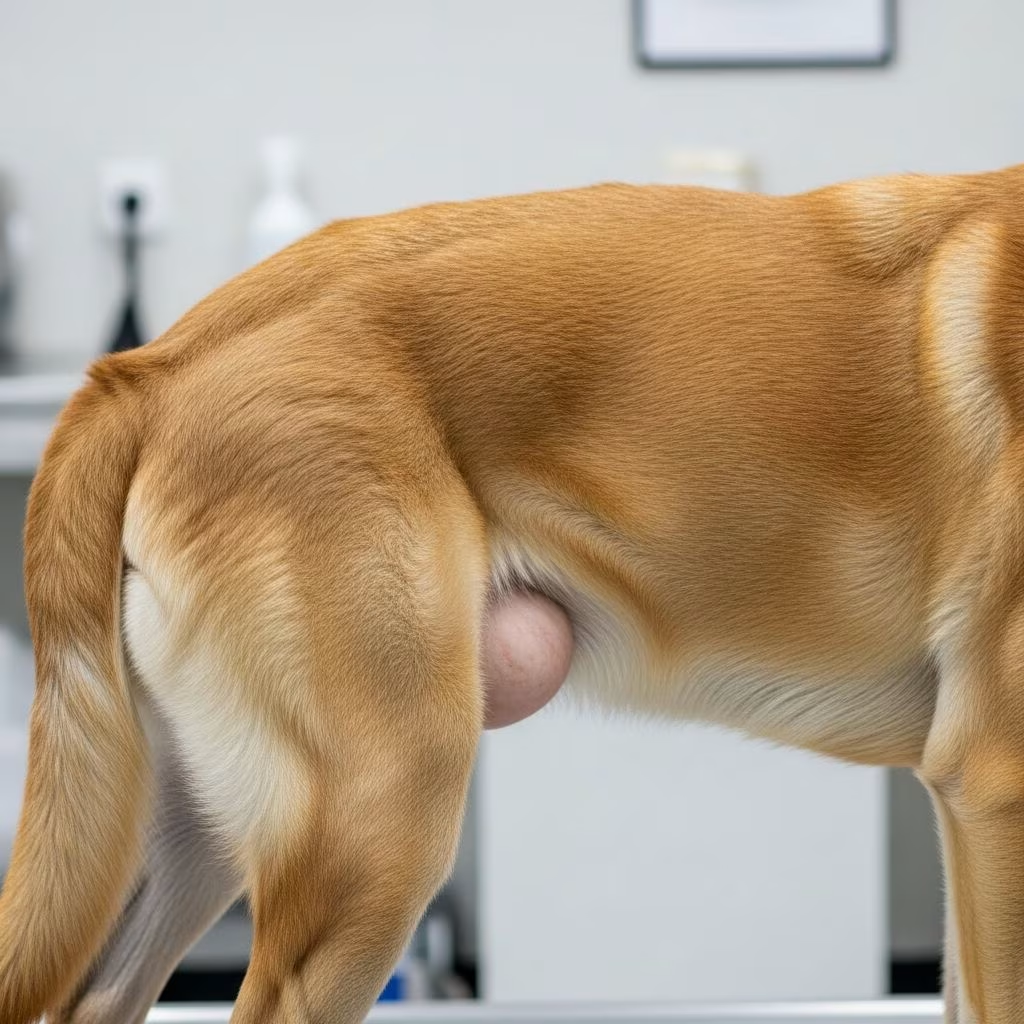
Understanding the Anatomical Basis of Perineal Hernias
A perineal hernia develops when the muscular structures that form the pelvic floor deteriorate or fail to maintain their structural integrity. Specifically, the levator ani and coccygeus muscles, which are responsible for supporting pelvic and abdominal organs, weaken or separate. This muscular failure creates an opening through which internal tissues can protrude into the perineal region—the area between the anus and genitals. When this happens, various abdominal and pelvic structures, including fat, fluid, the rectum, urinary bladder, and sometimes portions of the small intestine, can herniate through the defect.
The exact cause of perineal hernia formation in adult dogs remains incompletely understood, though the condition can occasionally occur congenitally in puppies. Several risk factors have been identified through clinical observation and research. The condition predominantly affects older, male dogs, with intact (non-castrated) males showing significantly higher incidence rates compared to their neutered counterparts. Chronic straining from conditions that impede normal defecation or urination may contribute to progressive weakening of the pelvic diaphragm. Additionally, chronic respiratory disease causing recurrent coughing could theoretically increase intra-abdominal pressure and contribute to muscular deterioration.
Recognizing the Clinical Presentation
Dogs with perineal hernias typically present with a constellation of clinical signs that vary in severity depending on which structures have herniated and the degree of compromise to those tissues. Understanding these manifestations helps pet owners recognize when professional veterinary evaluation is necessary.
Gastrointestinal Symptoms
The most common presenting complaint involves difficulty with normal bowel function. Affected dogs frequently strain excessively during defecation, a condition known as tenesmus. This straining occurs because the herniated rectum loses the supportive fascia and muscular structures that normally facilitate efficient stool passage. As the rectum becomes stretched and dilated due to herniation, normal peristaltic function is compromised. Constipation and obstipation (the inability to defecate) are frequent consequences, with fecal material accumulating proximal to the affected rectum. Dyschezia, defined as painful or difficult defecation, may cause affected dogs to spend extended periods in the squatting position with minimal productive effort.
Physical Manifestations
One of the most visible signs is perineal swelling, typically appearing as a soft, fluctuant bulge on one or both sides of the anus, lateral to the anal opening. This swelling results from entrapment of abdominal fat and fluid accumulation within the hernial sac. The swelling may vary in size depending on the dog’s posture and activity level, and it is usually non-painful upon examination. Some owners notice altered tail positioning or carriage, with affected dogs holding their tails lower than normal or exhibiting reduced tail mobility.
Urinary and Secondary Complications
When the urinary bladder herniates into the perineal space, more serious complications develop. Dogs experience stranguria (painful urination) or dysuria (difficult urination), and urine output may be severely reduced or absent. In acute cases where the herniated bladder becomes retroflexed (bent backward), complete urinary obstruction can occur, preventing normal micturition and leading to rapid accumulation of urine and potentially life-threatening uremia. Additional signs may include vomiting, flatulence, fecal incontinence, loss of appetite, and lethargy. If portions of the small intestine become trapped and strangulated, with compromised blood supply, the dog may experience acute abdominal pain, depression, and signs of shock.
Diagnostic Approach and Confirmation
Diagnosis of perineal hernia relies primarily on thorough history taking and physical examination. A veterinarian will palpate the perineal region to identify the characteristic soft swelling. The hernia often contains liquid, which may be detected through ballottement—a technique where gentle pressure elicits movement of contained fluid. Assessment of bladder position and content is crucial, particularly when urinary signs are present. If the hernia contains a full, distended bladder, palpation may reveal a tighter, more resilient consistency rather than the fluid wave typical of fat and fluid herniation.
Radiographic imaging provides valuable information about which tissues have herniated and the severity of displacement. Radiographs taken in lateral and ventrodorsal views can demonstrate retroflexion of the bladder, rectal deviation, and the presence of intestinal loops within the hernial defect. Ultrasonographic examination may further clarify the contents of the hernial sac and assess for any complications. In cases where urinary obstruction is suspected, urinalysis and serum biochemistry (to check for elevated creatinine and potassium indicating post-renal uremia) help guide emergency intervention. Peritoneocentesis—aspiration of peritoneal fluid—may be performed if bladder rupture or peritonitis is suspected. Some dogs with perineal hernias also have concurrent inguinal hernias, which should be identified during the diagnostic workup to ensure comprehensive surgical planning.
Medical Management and Conservative Approaches
In dogs with uncomplicated perineal hernias that do not involve bladder entrapment or intestinal strangulation, short-term conservative management may temporarily alleviate clinical signs. However, it is important to emphasize that medical management alone does not resolve the underlying anatomical defect and carries significant risk if complications develop unexpectedly.
Stool softeners and dietary modifications are commonly recommended components of conservative care. A diet high in fiber, often provided as a canned formulation, helps promote regular bowel movements and reduces excessive straining. Stool softeners decrease fecal consistency, making defecation easier and reducing the pressure increases associated with tenesmus. Pain management with appropriate analgesics may help reduce straining behavior. However, long-term reliance on medical management is contraindicated because life-threatening visceral entrapment and strangulation can occur suddenly, transforming a stable condition into an emergency requiring immediate surgical intervention.
Surgical Treatment: The Definitive Solution
Surgical hernia repair, known as herniorrhaphy, is the standard and recommended treatment for all perineal hernias. Emergency surgery is indicated immediately when the bladder has herniated and caused urinary obstruction, or when loops of intestine are strangulated with compromised blood supply. In these acute scenarios, delay in surgical intervention risks organ loss and potentially fatal complications.
Preoperative Considerations
Before herniorrhaphy, the surgeon assesses whether the urinary bladder is trapped within the hernial sac. If urinary obstruction is present and the hernia cannot be reduced manually, emergency catheterization is performed to decompress the bladder and relieve urinary pressure. A urinary catheter may remain indwelling postoperatively to ensure urethral patency and prevent recurrence of obstruction. In cases where catheterization is not immediately feasible, cystocentesis (needle aspiration directly from the bladder) provides emergency urine removal.
Surgical Technique and Reconstruction
The surgical approach to perineal hernia repair involves careful dissection to identify and separate the hernial contents from surrounding tissues and fibrous attachments. The surgeon precisely identifies the muscles forming the hernia defect—primarily the levator ani and coccygeus—along with important neurovascular structures including the internal pudendal artery and vein, the pudendal nerve, and the caudal rectal vessels and nerve. The sacrotuberous ligament, which plays a role in pelvic support, is also carefully preserved or incorporated into the repair.
One widely used technique involves elevating and transposing the internal obturator muscle from the pelvic floor to create a new pelvic diaphragm and reinforce the hernial repair. This muscle flap provides additional structural support to the repaired defect. The hernia is maintained in reduction by packing the defect with moistened, tagged sponges during the dissection and identification phase. Once the anatomy is clearly defined, suturing techniques restore the anatomical integrity of the pelvic diaphragm. In severe cases with extensive herniation, the abdomen may also need to be opened to allow surgical fusion of the rectum to the abdominal wall, permanently repositioning it out of the hernial sac.
Castration is performed during the same surgical procedure, typically through a median perineal incision. Castration significantly reduces the likelihood of hernia recurrence. Studies demonstrate that non-castrated dogs have recurrence rates approximately 2.7 times higher than castrated dogs, making this concurrent procedure an important component of successful repair.
Postoperative Tissue Biopsy
During surgery, any abnormal structures encountered within the hernial sac—such as the prostate or any masses—should be biopsied to rule out neoplasia or other pathology that might have contributed to the herniation or require separate treatment.
Postoperative Management and Recovery
Successful recovery from perineal hernia repair requires careful attention to pain control, wound management, dietary modifications, and activity restriction. The postoperative period is critical in determining whether the repair will heal properly or develop complications.
Pain Management and Hemorrhage Control
Analgesic medications are administered as necessary to minimize straining and reduce the risk of rectal prolapse, a complication where the rectum protrudes through the anus due to excessive tenesmus. Cold compresses applied immediately after surgery and repeated two to three times daily for 15 to 20 minutes during the first 48 to 72 hours help minimize hemorrhage and inflammatory swelling. After the initial 48 to 72-hour period, warm compresses applied two to three times daily for similar durations promote blood flow, reduce swelling, and alleviate perianal irritation.
Infection Monitoring and Systemic Treatment
Dogs receive broad-spectrum antibiotics during hospitalization and may continue on antimicrobial therapy at home. The surgical site should be monitored daily for signs of infection, including redness, increased pain, swelling, or discharge. Rectal prolapse, should it occur, is managed through careful reduction and placement of a purse-string suture around the anus to maintain proper anatomical position. Recurrent prolapse may necessitate a colopex procedure, which surgically anchors the colon to the abdominal wall to prevent future prolapse.
Dietary Support and Bowel Management
A high-fiber canned diet is provided to support normal bowel function while avoiding excessive straining. Stool softeners are continued for one to two months to maintain soft stool consistency and reduce defecation-related discomfort. This dietary approach is essential because painful defecation can lead to straining and constipation, creating a counterproductive cycle that stresses the surgical repair.
Activity Restriction
Dogs should be kept in a calm, quiet environment for the first two weeks following surgery to minimize activity that might increase intra-abdominal pressure or stress the surgical repair. Restricted exercise during this critical healing period significantly improves outcomes. Gradually increasing activity is permitted as healing progresses.
Prognosis and Long-Term Outcomes
Overall, approximately 85% of dogs undergoing surgical repair for perineal hernia experience successful outcomes with resolution of clinical signs and maintenance of the repair. However, the prognosis is guarded due to the significant risk of complications and recurrence.
Recurrence rates vary considerably in the literature, ranging from 10% to 46% depending on surgical technique, surgeon experience, and adherence to postoperative protocols. Complications beyond simple recurrence include infection, rectocutaneous fistula formation (abnormal communication between rectum and skin), anal sac fistulas, sciatic and pudendal nerve entrapment causing neurological dysfunction, and rectal prolapse. Castration during herniorrhaphy significantly improves recurrence rates and is strongly recommended as standard practice.
Frequently Asked Questions About Canine Perineal Hernias
Can perineal hernias be prevented?
While the exact etiology remains unclear, castrating male dogs before middle age may reduce the risk of developing this condition. Early intervention for conditions causing chronic straining, such as managing constipation or respiratory disease, may also help preserve pelvic floor integrity.
Is surgery always necessary?
Surgical repair is the only definitive treatment. Conservative management may provide temporary symptomatic relief but does not address the underlying defect and risks sudden life-threatening complications. Surgery should not be delayed once diagnosis is confirmed unless the dog’s overall health status requires stabilization first.
What happens if surgery is not performed?
Without surgical intervention, progressive herniation occurs, clinical signs worsen, and the risk of acute, life-threatening complications increases substantially. Bladder entrapment or intestinal strangulation can develop rapidly and may prove fatal without emergency surgical rescue.
How long is recovery after hernia repair?
Most dogs show significant clinical improvement within weeks of surgery. Complete tissue healing and return to normal activity typically requires two to three months. Stool softeners are usually continued for one to two months postoperatively.
References
- Perineal Hernia in Dogs — Merck Veterinary Manual. 2024. https://www.merckvetmanual.com/digestive-system/diseases-of-the-rectum-and-anus/perineal-hernia-in-dogs
- Perineal Hernia — American College of Veterinary Surgeons. 2024. https://www.acvs.org/small-animal/perineal-hernias/
- Perineal Hernia – Signs, Diagnosis and Treatment — Seven Oaks Pet Hospital. 2024. https://www.sevenoakspet.com/blog/perineal-hernia/
- Perineal Hernia — BluePearl Pet Hospital. 2024. https://bluepearlvet.com/medical-articles-for-pet-owners/perineal-hernia/
- Treatment for Hernias in Dogs — Zoetis Petcare. 2024. https://www.zoetispetcare.com/blog/article/treatment-hernia-dogs
- Perineal Hernia — Swift Referrals. 2024. https://www.swiftreferrals.co.uk/what-we-do/soft-tissue-surgery/perineal-hernia



