

Patent Ductus Arteriosus in Dogs: Understanding a Critical Congenital Heart Condition
Patent Ductus Arteriosus (PDA) represents one of the most common congenital heart defects affecting dogs, particularly small and toy breed dogs. This hereditary cardiac condition occurs when a blood vessel called the ductus arteriosus fails to close properly after birth, creating an abnormal connection between two major blood vessels in the heart. Understanding PDA, its symptoms, treatment options, and prognosis is essential for dog owners whose pets may be affected by this serious condition.
What is Patent Ductus Arteriosus?
The ductus arteriosus is a normal blood vessel that exists in puppies before birth. During fetal development, this vessel allows blood to bypass the lungs because the fetus receives oxygen through the mother’s placenta rather than through its own lungs. Normally, this vessel closes within the first few days or weeks after a puppy is born, once the puppy begins breathing air and no longer needs this bypass.
In dogs with PDA, however, this vessel fails to close properly. This creates an abnormal shunt that allows blood to flow continuously from the aorta (the main artery carrying blood away from the heart) directly into the pulmonary artery (which carries blood to the lungs). This abnormal blood flow bypasses the normal circulatory pathway and places additional strain on the heart and lungs.
Early epidemiological studies have suggested a genetic component to the occurrence of PDA, meaning that susceptibility to this condition can be inherited from parent dogs to their offspring. Small and toy breed dogs appear to be predisposed to this condition, although dogs and cats of any breed and size can be affected.
Signs and Symptoms of PDA
The clinical presentation of PDA varies depending on the size of the defect and the severity of the abnormal blood flow through the shunt. Dogs with small PDA defects may not show any obvious signs of disease initially.
Early and Mild Symptoms
As the PDA increases in size and more blood is shunted through it, dogs may develop mild clinical signs including:
- Difficulty breathing or rapid breathing
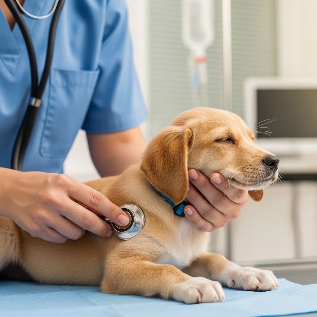
- A loud, continuous heart murmur audible with a stethoscope
- Abnormal pulses, often described as “bounding” or hyperkinetic
- Exercise intolerance
- Stunted growth or smaller than average size
- Excessive sleeping or lethargy
- Being thinner than average for their age
Advanced Symptoms
As blood continues to shunt through the PDA, it causes irreversible changes in the heart muscle and eventually may lead to congestive heart failure. If the PDA is significant enough to cause congestive heart failure, additional signs may develop including increased lung sounds when listened to with a stethoscope, heart arrhythmias (abnormal heart beats), accumulation of fluid in the lungs and/or abdomen, severe activity intolerance, coughing, dyspnea (difficulty breathing), and exertional tachypnea (rapid breathing with exercise).
Understanding Reverse PDA
In some severe cases, reverse PDA can develop. This occurs when the shunt is very large and the pressure in the pulmonary circulation continues to increase until it exceeds the pressure in the aorta. When this happens, the direction of blood flow reverses, so that blood returning from the body shortcuts through the aorta before going through the lungs to receive oxygen.
Reverse PDA is a critical condition characterized by severe and debilitating signs including:
- Disappearance of the heart murmur
- Abnormal heartbeats
- Cyanosis (blueness of the foot pads, particularly on the hind legs)
- Collapse of the hind legs during exercise
- Severe weakness or lethargy
Diagnosis of PDA
Diagnosis of PDA typically begins when a veterinarian detects a loud, continuous heart murmur during a routine physical examination. The distinctive murmur associated with PDA is often one of the first indicators of the condition. To confirm the diagnosis and assess the severity of the defect, veterinarians may recommend additional diagnostic tests including chest radiographs (X-rays), echocardiography (heart ultrasound), electrocardiography (ECG), and in some cases, cardiac catheterization. These diagnostic tools help determine the size of the PDA, the direction of blood flow, and whether any complications such as pulmonary edema or congestive heart failure have developed.
Treatment Options for PDA
The goal of treatment for a forward-flowing PDA is to stop the blood flowing through the abnormal shunt. Your veterinarian will refer you to a veterinary cardiovascular surgeon who will determine the optimal treatment approach for your dog’s unique situation. Treatment decisions depend on several factors including the dog’s size, the size of the PDA, and whether any irreversible heart damage has already occurred.
Surgical Ligation
Traditional surgical treatment involves ligation (tying off) of the ductus arteriosus during open-chest heart surgery. This procedure requires an incision between the ribs to access the heart and the abnormal vessel. The PDA is identified, gently isolated, and closed using permanent suture material. This remains an effective and proven treatment option, particularly for smaller dogs where the anatomy may not be suitable for minimally invasive procedures. Early intervention with surgical ligation has proven curative if performed by 6 months of age.
Minimally Invasive Catheterization
In many cases, less invasive occlusion of the PDA is now possible using modern interventional cardiology techniques. This procedure involves placing a long catheter in a hind leg and passing it into the abnormal vessel using fluoroscopic guidance. A specialized device called an Amplatzer ductal occluder is then deployed to stop the abnormal blood flow through the PDA. This minimally invasive approach has the advantage of being less traumatic than open-chest surgery, requiring no incisions and resulting in faster recovery times.
Not all PDAs can be treated using minimally invasive techniques, as patient size, anatomy, and the size of the PDA are paramount considerations. The cardiologist attending your dog’s case will advise on the best option for your specific situation.
Pre-surgical Stabilization
If the dog shows signs of congestive heart failure, pre-surgical stabilization will be necessary before correction of the PDA can be performed. If pulmonary edema (fluid in the lungs) is visible on radiographs, it is essential to treat with diuretics for 24 to 48 hours before surgery or coil embolization. This medical management helps stabilize the patient and reduces the risk of complications during the definitive surgical procedure.
Treatment for Reverse PDA
Unfortunately, there is no surgical treatment for a reverse PDA. Medical treatment will help manage the symptoms through heart medications, but a cure is not possible for dogs with a reverse PDA, and the condition will inevitably be fatal. However, if the signs of disease can be controlled with medication, the dog may live comfortably for several years with appropriate medical management and careful monitoring.
Success Rates and Outcomes
The prognosis for PDA depends significantly on when treatment is initiated and whether irreversible heart damage has already occurred. If your pet receives treatment before experiencing heart failure, the success rate associated with surgery is quite high, and the majority of treated dogs can return to normal life with excellent long-term prognosis.
Both standard surgical and minimally invasive transcatheter techniques can successfully treat the condition as long as it hasn’t progressed to a reverse PDA. If irreversible heart damage has already occurred, your dog might need to remain on heart medication in the future, but appropriate treatment can still significantly improve quality of life.
In contrast, the prognosis for untreated PDA is poor. Research has shown that 64% of untreated dogs died within 1 year of examination. Additionally, 65% of dogs will die from the development of congestive heart failure by 1 year of age if occlusion or ligation of the PDA is not performed.
Importance of Early Detection and Treatment
Surgical repair should be performed as soon as possible once a diagnosis is confirmed. The longer the surgery is delayed, the more likely it is that irreversible heart damage will occur. Early intervention is critical because the continuous abnormal shunt places progressive strain on the heart muscle, eventually leading to permanent changes that cannot be reversed even after the PDA is corrected.
If left untreated, most dogs will develop congestive heart failure and experience serious complications including labored breathing, cough, exercise intolerance, fatigue, and ultimately death. Without proper management, death may occur sometimes quickly or after a few days once congestive heart failure symptoms become severe.
Post-operative Considerations
After successful surgical or catheter-based treatment of PDA, most dogs can return to normal activity and life expectancy. During the recovery period, your veterinarian will provide specific instructions regarding activity restriction, medication administration, and follow-up appointments. In cases where irreversible heart damage has occurred prior to treatment, ongoing cardiac medication may be necessary to manage residual heart dysfunction.
Frequently Asked Questions About PDA in Dogs
Q: Is PDA a hereditary condition?
A: Early epidemiological studies have suggested a genetic component to the occurrence of PDA, indicating that susceptibility can be inherited. Small and toy breed dogs appear to be predisposed to this condition, although dogs of any breed and size can be affected.
Q: What is the difference between forward and reverse PDA?
A: In forward PDA, blood flows from the aorta to the pulmonary artery through the abnormal vessel. In reverse PDA, the pressure becomes so high that blood flow reverses, causing blood to bypass the lungs and resulting in severe oxygen deficiency. Reverse PDA is more serious and has a poorer prognosis.
Q: Can medical management alone cure PDA?
A: Medical management has not proven to be effective in treating PDA in dogs. Only surgical ligation or catheter-based occlusion can close the abnormal vessel. Medical treatment is only used to manage symptoms and stabilize patients before surgery or in cases of reverse PDA where surgery is not an option.
Q: What is the recovery time after PDA surgery?
A: Recovery time varies depending on whether traditional surgery or minimally invasive catheterization was performed. Minimally invasive procedures typically have faster recovery times, while traditional open-chest surgery may require a longer recovery period. Your veterinary surgeon will provide specific post-operative instructions for your dog.
Q: What is the long-term prognosis after successful PDA treatment?
A: If treatment is performed before irreversible heart damage occurs, the long-term prognosis is excellent. Most dogs can return to normal life and activities with a normal life expectancy. However, if heart damage has already occurred, ongoing medication may be necessary.
Q: Why is early diagnosis and treatment so important for PDA?
A: Early treatment is critical because the continuous abnormal shunt causes progressive and irreversible damage to the heart muscle. The longer treatment is delayed, the higher the risk of permanent heart damage that cannot be reversed even after the PDA is corrected.
References
- Patent Ductus Arteriosus in the Canine Patient — Mississippi State University College of Veterinary Medicine. 2017. https://www.vetmed.msstate.edu
- Patent Ductus Arteriosus (PDA) in Dogs — CVCA Cardiac Care for Pets. https://www.cvcavets.com
- Cardiac Shunts in Animals — Merck Veterinary Manual. https://www.merckvetmanual.com
- Patent Ductus Arteriosus (PDA) in Dogs — University of Florida Small Animal Hospital. https://smallanimal.vethospital.ufl.edu
- Patent Ductus Arteriosus (PDA) Fact Sheet — Veterinary Specialists UK. https://www.vetspecialists.co.uk
- Patent Ductus Arteriosus (PDA) in Dogs — VRCC Veterinary Emergency Hospital, Englewood. https://www.vrcc.com



