

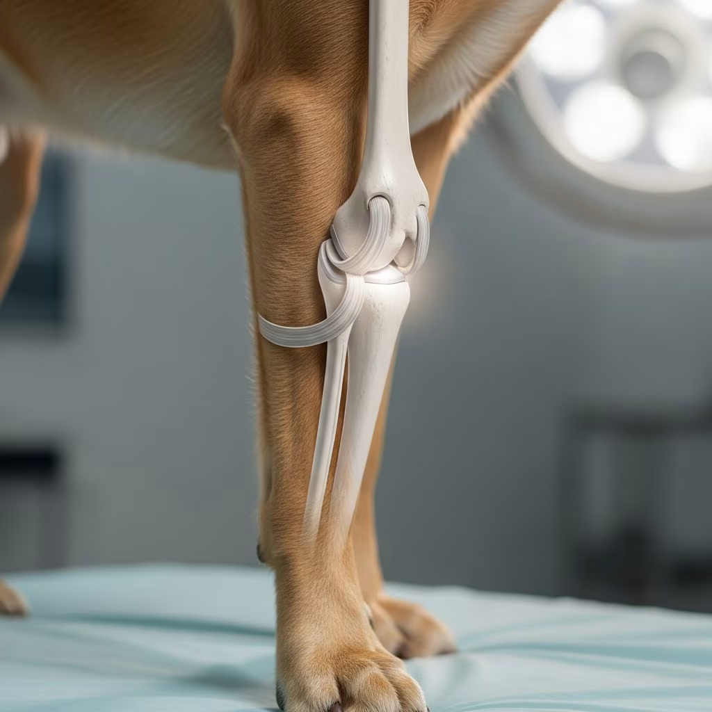
Patellar luxation occurs when a dog’s kneecap slips out of its normal position in the groove of the femur, leading to discomfort, abnormal movement, and potential long-term joint issues. This condition is particularly prevalent in small breeds but can affect dogs of various sizes, requiring prompt recognition and tailored interventions to maintain quality of life.
Understanding the Anatomy and Mechanism of Patellar Luxation
The patella, or kneecap, functions as a critical component of the canine stifle joint, which is equivalent to a human knee. It sits within a trochlear groove on the femur and is held in place by ligaments and surrounding muscles. When luxation happens, the patella displaces medially (inward) or laterally (outward), most commonly medially in dogs. This misalignment disrupts normal joint mechanics, causing instability and wear on cartilage over time.
Several anatomical factors contribute to this slippage. Shallow trochlear grooves, misaligned tibial tuberosities, or lax joint capsules can predispose dogs to luxation. Trauma from falls or rough play may trigger acute episodes, but congenital deformities are more common culprits, explaining the hereditary nature in certain lineages.
Common Causes and Risk Factors
Genetic predisposition tops the list of causes, with small breeds like Chihuahuas, Pomeranians, Maltese, and Yorkshire Terriers showing higher incidence rates due to inherited shallow grooves and ligament weaknesses. Larger breeds such as Labrador Retrievers can also develop it, often bilaterally.
- Breed susceptibility: Toy and miniature varieties face elevated risks from selective breeding emphasizing aesthetics over structural integrity.
- Trauma: Injuries from jumps, slips, or collisions can displace the patella acutely.
- Obesity: Excess weight strains joints, exacerbating instability.
- Developmental issues: Rapid growth in puppies may lead to conformational flaws.
Preventive measures focus on weight control, controlled exercise, and early screening in at-risk puppies through veterinary orthopedic exams.
Recognizing the Signs: Symptoms to Watch For
Early detection hinges on observing subtle behavioral and gait changes. Dogs often display intermittent lameness, where they hold up a hind leg briefly before resuming normal walking, resembling a skip or hop.
| Symptom | Description | Frequency |
|---|---|---|
| Skipping gait | Sudden leg lift during walk or run, as if kicking to reposition kneecap | Common in early stages |
| Limping | Intermittent favoring of hind leg, worse after activity | Progresses with severity |
| Pain indicators | Yelps, reluctance to jump, or stiffness upon rising | Variable, increases in advanced cases |
| Postural changes | Bow-legged stance, hunched back, or abnormal hind leg alignment | Chronic sign |
| Joint sounds | Cracking or popping from knee area | Occasional |
Owners might palpate a loose kneecap during gentle exams, but professional diagnosis via physical manipulation and X-rays confirms the issue and rules out fractures or ligament tears.
Grading the Severity: From Mild to Critical
Veterinarians classify patellar luxation into four grades based on stability and manual repositioning ease, guiding treatment decisions.
- Grade I: Patella slips out manually but snaps back spontaneously; minimal symptoms, often asymptomatic.
- Grade II: Occasional spontaneous luxation requiring manual replacement; intermittent lameness prompts monitoring or conservative care.
- Grade III: Patella mostly luxated, reducible manually with effort; persistent lameness and muscle atrophy common.
- Grade IV: Permanent dislocation, irreducible; severe pain, deformity, and rapid joint degeneration necessitate intervention.
Progression from lower to higher grades occurs without management, underscoring early veterinary consultation.
Non-Surgical Management Strategies
For Grades I and II, conservative approaches effectively control symptoms and delay progression. These include multimodal care emphasizing rest, medication, and rehabilitation.
- Weight management: Reducing body fat alleviates joint stress; prescription diets aid compliance.
- Medications: NSAIDs like carprofen or grapiprant reduce inflammation and pain; never use human drugs without vet approval.
- Supplements: Glucosamine, chondroitin, and omega-3s support cartilage repair and lubrication.
- Exercise modification: Short leash walks, no stairs or jumping; hydrotherapy builds muscle without impact.
- Physical therapy: Targeted exercises strengthen quadriceps and stabilizers; professional sessions optimize outcomes.
Improvement may take weeks, with periodic flare-ups managed by adjusting protocols. Braces offer additional stability in select cases.
Surgical Interventions for Advanced Cases
Grades III and IV, or failed conservative treatments, warrant surgery to realign anatomy and restore function. Success rates exceed 90% in restoring mobility when performed by specialists.
Common techniques include:
- Trochlear groove deepening: Resects bone to create a deeper femoral slot (wedge or block recession).
- Tibial tuberosity transposition: Repositions shinbone attachment for better patellar tracking.
- Capsular adjustments: Tightens lax tissues and releases tight ones (imbrication and release).
- Fabellar sutures: Temporary stabilization during healing.
Procedures last 1-2 hours per knee, often bilateral. Risks like infection or implant failure are low with proper aftercare.
Post-Treatment Recovery and Long-Term Care
Conservative recovery spans 3-6 weeks of restricted activity, while surgical healing demands 8-12 weeks crate rest, gradual rehab, and frequent vet checks.
- Immediate post-op: E-collar, pain meds, and sling support for walking.
- Rehab phase: Passive range-of-motion, underwater treadmill, and balancing exercises.
- Lifelong maintenance: Annual exams, supplements, and low-impact activities like swimming.
Prognosis excels with compliance; most dogs regain near-normal function, though monitoring prevents recurrence.
Prevention Tips for At-Risk Dogs
Proactive steps mitigate risks:
- Select breeders screening for orthopedic health.
- Maintain ideal weight via portion control and activity.
- Avoid high-impact play in puppies.
- Incorporate joint-friendly exercises early.
Regular vet wellness exams catch issues pre-symptomatically.
Frequently Asked Questions (FAQs)
Can patellar luxation heal on its own?
Mild Grade I cases may stabilize with management, but progression is common without intervention.
Is surgery always necessary?
No, low-grade cases thrive on conservative care; surgery reserved for severe or unresponsive instances.
How much does surgery cost?
Varies by location and complexity, typically $1,500-$5,000 per knee; pet insurance offsets expenses.
What breeds are most affected?
Small breeds like Pomeranians, Pugs, and Shih Tzus; also seen in Boston Terriers and Cavaliers.
Can both knees be affected?
Yes, bilateral luxation occurs in up to 50% of cases, often requiring staged surgeries.
Choosing the Right Specialist
Board-certified veterinary surgeons or orthopedists offer best outcomes. Seek clinics with rehab facilities for comprehensive care.
References
- Luxating Patella in Dogs: Prevention, Symptoms, Treatment, and Surgery — Animal Outpatient Surgery. 2023. https://animaloutpatientsurgery.com/blog/luxating-patella-in-dogs-prevention-symptoms-treatment-and-surgery/
- Luxating Patella in Dogs: What Is It, and How Is It Treated? — PetMD. 2024-02-15. https://www.petmd.com/dog/conditions/musculoskeletal/luxating-patella-in-dogs
- Luxating Patella in Dogs — Dogwood Veterinary Hospital. 2023. https://dogwoodvet.com/blog/luxating-patella-in-dogs/
- Luxating Patella in Dogs — VCA Animal Hospitals. 2024. https://vcahospitals.com/know-your-pet/luxating-patella-in-dogs
- Luxating Patella in Dogs: Symptoms, Treatment Options & Prognosis — Bond Vet. 2023. https://bondvet.com/blog/luxating-patella-dog
- Luxating Patella in Dogs: Causes, Symptoms, & Treatment — TPLO Info. 2024. https://tploinfo.com/blog/luxating-patella-in-dogs-causes-symptoms-treatment/



