

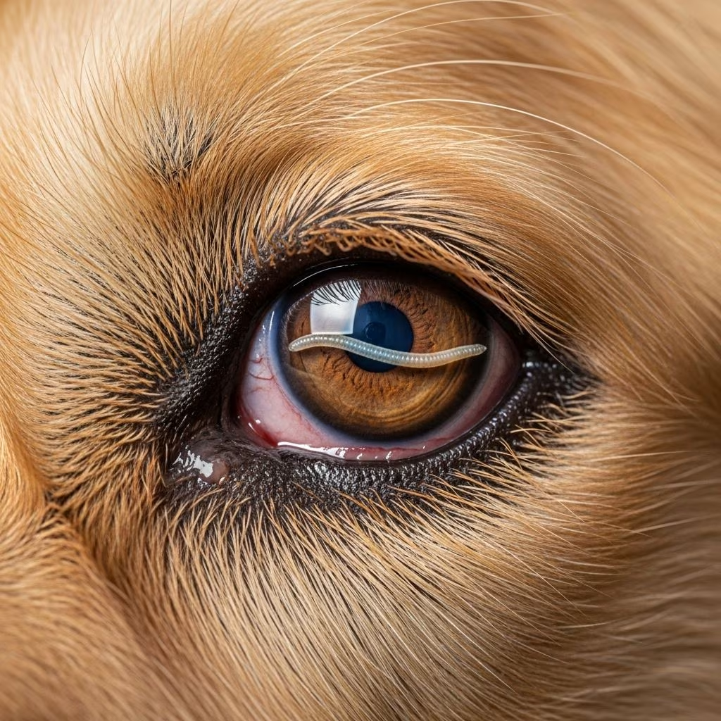
Parasitic infections affecting the eyes represent a significant but often overlooked health concern for pet owners. These infections, caused by nematode parasites primarily from the genus Thelazia, can affect both dogs and cats, creating considerable discomfort and potentially leading to vision loss if left untreated. Understanding the nature of these infections, their clinical presentations, and available treatment modalities is essential for anyone responsible for companion animal care.
The Nature of Ocular Parasitic Infections
Parasitic eye infections occur when nematode worms establish themselves within the ocular tissues and associated structures of affected animals. These parasites are spirurid nematodes that have adapted to survive in the unique environment of the eye and its surrounding tissues. The infection is mediated through insect vectors, primarily fruit flies, which transmit infective larvae to susceptible hosts during feeding activities.
The parasites themselves are microscopic to small organisms, measuring approximately 10 to 15 millimeters in length, appearing white to cream in coloration. Once introduced into the eye, these organisms can reside in multiple ocular locations, making complete removal and treatment challenging. The parasites prefer to inhabit the conjunctival tissues, the protective membranes lining the eye, but can also migrate to the nictitating membrane (third eyelid), lacrimal glands, and corneal surfaces.
Geographic Distribution and Risk Factors
Historically, these infections were considered primarily a concern in warmer climates with high populations of insect vectors. However, recent epidemiological trends suggest a geographic expansion of cases. Notable increases have been documented in temperate regions such as the Hudson Valley, where climate changes have contributed to extended seasons favorable for fly populations.
Several factors influence transmission risk:
- Outdoor exposure duration and frequency
- Environmental temperature and humidity patterns
- Population density of competent insect vectors
- Individual animal susceptibility and immune status
- Lapses in preventative medication administration
Pets with increased outdoor activities face heightened exposure, particularly during seasons when insect vector populations peak. Additionally, imported animals from endemic regions may introduce infections to previously unaffected areas.
Clinical Manifestations and Disease Progression
The clinical presentation of parasitic eye infections spans a spectrum from subclinical carriage to severe pathological conditions. Infected animals may exhibit minimal signs initially, with disease progression influenced by parasite burden, individual immune response, and duration of infection.
Common clinical signs include:
- Excessive eye discharge (epiphora)
- Conjunctival inflammation and redness
- Ocular itching and discomfort
- Swelling of conjunctival tissues (chemosis)
- Cloudiness or opacity of the cornea
- Increased blinking frequency
- Light sensitivity
In advanced cases, severe complications develop. Corneal ulceration represents one of the most concerning sequelae, involving loss of the protective epithelial layer covering the cornea. These ulcerations can progress to secondary bacterial infections, potentially resulting in permanent scarring, vision impairment, and in untreated cases, complete blindness.
Diagnostic Approaches for Veterinary Professionals
Veterinary diagnosis of parasitic eye infections typically relies on direct visualization of parasites, making ophthalmologic examination central to diagnostic confirmation. The presence of visible nematodes on the conjunctival surface or under the eyelids provides definitive diagnosis.
Examination Techniques:
Comprehensive eye examination may require topical anesthesia to facilitate visualization of structures beneath the eyelids. Specialized equipment including slit lamp biomicroscopy allows detailed examination of anterior eye structures, enabling identification of parasites, assessment of corneal integrity, and evaluation of inflammatory changes.
Additional diagnostic modalities may include:
- Schirmer tear tests to quantify tear production
- Fluorescein staining to identify corneal epithelial defects
- Intraocular pressure measurement to assess glaucoma risk
- Nasolacrimal duct flushing to remove parasites and secretions
Parasitological confirmation through submission of removed organisms to specialized diagnostic laboratories ensures accurate identification and guides treatment decisions.
Treatment Strategies: A Multimodal Approach
Effective management of parasitic eye infections typically requires integration of multiple therapeutic modalities, including mechanical removal, pharmacological intervention, and supportive care.
Mechanical Extraction
Physical removal of visible parasites remains the primary treatment approach in most clinical cases. Under topical anesthesia or systemic sedation, veterinarians carefully extract worms using atraumatic forceps or gentle saline irrigation techniques. This direct approach provides immediate parasite burden reduction and allows for specimen collection for diagnostic confirmation.
Mechanical removal offers several advantages: immediate symptom relief, diagnostic confirmation through parasite examination, and reduced risk of medication-induced adverse effects. However, this approach requires professional expertise to minimize ocular tissue trauma during extraction procedures.
Antiparasitic Medications
Systemic antiparasitic agents provide parasiticide activity against ocular nematodes. Several medication classes demonstrate efficacy:
| Medication Class | Examples | Administration Route | Notes |
|---|---|---|---|
| Macrocyclic Lactones | Ivermectin, Moxidectin | Oral, Topical, Injectable | Commonly used in preventative formulations |
| Neonicotinoids | Imidacloprid | Topical | Often combined with moxidectin |
| Phenothiazine Derivatives | Selamectin | Topical | Alternative option for select cases |
Medication selection depends on case-specific factors including infection severity, presence of corneal complications, and individual animal tolerance profiles. Combined formulations containing both imidacloprid and moxidectin provide comprehensive antiparasitic coverage while addressing multiple parasite species.
Supportive Ocular Care
Topical and systemic medications address inflammatory sequelae of parasitic infection. Antibiotic ointments and drops prevent or treat secondary bacterial infections that frequently complicate corneal ulcerations. Anti-inflammatory medications, including topical corticosteroids and systemic nonsteroidal agents, reduce ocular inflammation and accelerate corneal healing.
Duration of supportive therapy typically spans 7-14 days post-extraction, with therapeutic response monitored through follow-up examinations. Complete corneal re-epithelialization requires confirmation through fluorescein staining before discontinuing topical medications.
Prevention: The Cornerstone of Disease Management
Preventing parasitic eye infections proves far more effective and cost-efficient than treating established infections. A comprehensive prevention strategy integrates pharmacological, behavioral, and environmental interventions.
Monthly Preventative Medications
Regular administration of heartworm preventatives containing moxidectin or other effective antiparasitic agents provides protection against ocular parasites. Consistent year-round administration proves critical, as even brief lapses in medication compliance create windows of vulnerability for infection.
These preventative medications operate through systemic circulation, achieving concentrations sufficient to kill parasitic larvae before they establish in ocular tissues. Monthly formulations align with heartworm prevention protocols, simplifying compliance for pet owners.
Behavioral and Environmental Modifications
Reducing exposure to insect vectors provides supplementary protection alongside pharmacological prevention:
- Limiting outdoor activities during peak insect season
- Avoiding areas with high fly concentrations
- Applying pet-safe insect repellents to susceptible animals
- Maintaining clean ocular and periocular areas
- Minimizing exposure during dawn and dusk hours when vectors are most active
These behavioral modifications prove particularly valuable in endemic regions and during seasons with elevated vector populations.
Prognosis and Long-Term Outcomes
The prognosis for parasitic eye infections depends significantly on timing of diagnosis and initiation of treatment. Animals receiving prompt veterinary intervention typically achieve complete clinical recovery without lasting visual impairment.
Early-stage infections with minimal corneal involvement generally respond excellently to treatment, with complete resolution within 7-14 days of initiating therapy. Follow-up examinations at 7-14 days post-treatment confirm parasite elimination and assess corneal healing progression.
Delayed diagnosis and treatment carry substantially worse prognoses. Chronic inflammation, extensive corneal ulceration, and secondary bacterial infections create permanent structural damage to ocular tissues. In severe untreated cases, permanent vision loss or blindness represents a significant risk. These complications underscore the importance of prompt veterinary evaluation when clinical signs suggestive of ocular disease develop.
Frequently Asked Questions
Can humans contract parasitic eye infections from infected pets?
While spirurid nematodes primarily affect companion animals, they can occasionally infect humans. However, transmission through direct contact with infected pets is extremely rare, and the parasite requires specific insect vector-mediated transmission.
How quickly do symptoms develop after parasitic exposure?
Clinical signs typically emerge 1-3 weeks following parasitic infection, though this timeline varies based on parasite burden and individual immune response. Some animals may remain subclinically infected with minimal signs for extended periods.
Is mechanical removal sufficient, or are medications always necessary?
While mechanical extraction addresses visible parasites, adjunctive antiparasitic medications eliminate larvae and prevent recurrence. Combining mechanical removal with antiparasitic therapy provides optimal outcomes.
What is the cost of treating parasitic eye infections?
Treatment costs vary based on infection severity, geographic location, and whether specialist ophthalmology referral is necessary. Uncomplicated cases with mechanical removal and topical medications cost considerably less than cases requiring specialist evaluation and management of corneal complications.
Can preventative medications be used year-round?
Yes, year-round monthly preventative administration is recommended, particularly in regions with extended transmission seasons. This approach provides consistent protection without requiring owners to determine seasonal risk periods.
References
- Three cases of imported eyeworm infection in dogs — National Center for Biotechnology Information (NCBI). 2017. https://pmc.ncbi.nlm.nih.gov/articles/PMC5738592/
- Eyeworm Infection in Dogs — PetMD. https://www.petmd.com/dog/conditions/eyes/eyeworm-infection-dogs
- Eye Worm in Dogs: Symptoms and Treatment — Blue Cross. https://www.bluecross.org.uk/advice/dog/health-and-injuries/eye-worm-danger-to-dogs
- Ocular Worms in Dogs and Cats: What You Need to Know — Middle Hope Veterinary Clinic. https://middlehopevet.com/news/ocular-worms-in-dogs-and-cats-what-you-need-to-know/
- How to Prevent Your Pet from Getting Eyeworms — Animal Emergency Care of New Jersey. https://www.aecofnj.com/blog/how-to-prevent-your-pet-from-getting-eyeworms
- DPDx – Thelaziasis — Centers for Disease Control and Prevention (CDC). https://www.cdc.gov/dpdx/thelaziasis/index.html



