

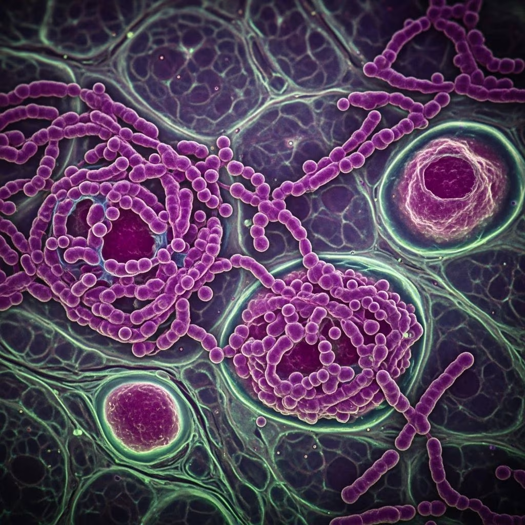
Nocardiosis represents a significant bacterial infection in various animal species, primarily caused by environmental bacteria from the Nocardia genus. These gram-positive, filamentous organisms thrive in soil and decaying vegetation, gaining entry through wounds or inhalation, leading to localized or systemic disease. Understanding this condition is crucial for timely intervention in veterinary practice.
Understanding the Pathogen Behind Nocardiosis
The Nocardia species are aerobic, branching bacteria partially acid-fast, commonly found in natural environments worldwide. Key species include Nocardia asteroides, Nocardia farcinica, and Nocardia veterana, each capable of causing opportunistic infections. In animals, infections often stem from traumatic injuries or respiratory exposure, with dissemination possible in vulnerable hosts.
These bacteria exhibit multidrug resistance, complicating therapy. For instance, N. farcinica isolates from canine cases have shown resistance to vancomycin and other agents. Predisposing factors include immunosuppression from medications like cyclosporine or prednisone, or concurrent viral infections such as canine distemper.
Species-Specific Manifestations of the Disease
Nocardiosis presents differently across animal classes, influenced by exposure routes and immune status.
- Dogs: Frequently develop disseminated forms with skin abscesses, lameness from limb lesions, pneumonia, and organ involvement like lungs, kidneys, and brain. Traumatic wounds serve as entry points, leading to sudden systemic illness.
- Cats: Respiratory and disseminated infections predominate, with abscesses in multiple organs and poor response to short-term treatments.
- Horses: Pulmonary nocardiosis causes severe pneumonia, pleuritis, and subcutaneous abscesses; systemic spread results in multi-organ abscessation and occasional abortion.
- Cattle and Other Livestock: Mastitis and pulmonary forms are noted, with chronic granulomatous lesions.
In dogs, cases often mimic other conditions like fungal infections or tumors, delaying diagnosis.
Recognizing Clinical Signs Early
Early detection hinges on identifying subtle initial symptoms that progress rapidly. Common indicators include:
- Respiratory distress: Coughing, labored breathing, nasal discharge, and pleural effusion.
- Cutaneous changes: Firm subcutaneous nodules, draining abscesses, especially near wounds or bite sites.
- Systemic effects: Lameness, fever, lethargy, neurological deficits from brain abscesses, and organ failure signs.
- Musculoskeletal issues: Osteomyelitis and joint involvement leading to pain and mobility loss.
In severe cases, pyothorax or pyogranulomatous lymphadenitis emerges, as seen in canine thoracic imaging with compressed bronchi and hypoxemia.
Diagnostic Approaches for Accurate Identification
Diagnosis combines clinical evaluation, imaging, cytology, and microbiology. Veterinarians start with:
| Method | Purpose | Key Findings |
|---|---|---|
| Cytology & Impression Smears | Initial screening | Branching filamentous bacteria amid inflammation |
| Culture & MALDI-TOF MS | Species identification | Rapid detection of N. farcinica or N. veterana |
| 16S rRNA Sequencing | Confirmation | Precise genotyping for resistance profiling |
| Imaging (X-rays, CT) | Lesion localization | Abscesses, lymphadenopathy, pulmonary infiltrates |
| Biopsy/Necropsy | Definitive pathology | Pyogranulomatous inflammation |
Cultures from blood, pus, or tissues are essential, though slow-growing. Advanced tools like MALDI-TOF accelerate identification over traditional sequencing.
Treatment Strategies and Challenges
Management requires prolonged antimicrobials (1-6 months), surgical intervention, and immunosuppression reversal where possible. Sulfonamides are first-line, but susceptibility testing guides choices due to resistance.
- Antibiotics: Trimethoprim-sulfamethoxazole, amikacin, or linezolid combinations; monitor for relapse.
- Surgery: Drainage of abscesses, debridement of wounds, thoracotomy for pyothorax.
- Supportive Care: Fluids, pain control, nutritional support.
Success stories include a dog with N. veterana bacteremia resolving after 6-8 weeks of therapy post-immunosuppressant cessation. However, mortality remains high in disseminated cases due to delayed diagnosis or financial limits.
Prevention and Control Measures
No vaccines exist, so focus on hygiene and risk reduction:
- Maintain clean environments, especially for housed animals.
- Treat wounds promptly and avoid trauma in high-risk groups.
- Minimize immunosuppressants; screen for underlying conditions.
- Early diagnostics in lameness or respiratory cases with trauma history.
In outbreaks, isolate cases and enhance biosecurity.
Prognosis Factors Influencing Outcomes
Prognosis varies by site and timeliness: cutaneous cases fare best (~100% cure in humans analogously), while disseminated forms have ~3-50% survival in vets. Positive factors include early surgery, targeted antibiotics, and immune competence. Poor indicators: CNS involvement, multidrug resistance, and comorbidities.
Emerging Research and Diagnostic Advances
Recent studies highlight MALDI-TOF’s role in speeding identification, vital for fastidious Nocardia. Outbreaks like N. farcinica in dogs underscore trauma’s role without overt immunosuppression. Future directions: resistance surveillance and novel therapies.
Frequently Asked Questions (FAQs)
What causes nocardiosis in pets?
Soil-dwelling Nocardia bacteria enter via wounds or lungs, thriving in immunocompromised animals.
Is nocardiosis contagious between animals?
No direct transmission; it’s environmentally acquired, not animal-to-animal.
How long does treatment last?
Typically 1-6 months of antibiotics, with monitoring to prevent relapse.
Can nocardiosis be fatal?
Yes, especially disseminated forms, but early intervention improves odds.
What breeds are most at risk?
No breed predisposition, but large dogs with outdoor exposure may encounter more.
This overview equips veterinarians and owners with knowledge to combat nocardiosis effectively, emphasizing vigilance and advanced diagnostics.
References
- Disseminated Nocardiosis Caused by Nocardia farcinica in Two Dogs — PMC. 2023. https://pmc.ncbi.nlm.nih.gov/articles/PMC9860694/
- Bacterial Infection (Nocardiosis) in Dogs — PetMD. Accessed 2026. https://www.petmd.com/dog/conditions/respiratory/c_dg_nocardiosis
- Successful Treatment of Disseminated Nocardiosis Caused by Nocardia veterana — PMC. 2018. https://pmc.ncbi.nlm.nih.gov/articles/PMC5787162/
- Nocardiosis in Animals — MSD Veterinary Manual. Accessed 2026. https://www.msdvetmanual.com/infectious-diseases/nocardiosis/nocardiosis-in-animals
- Disseminated Nocardiosis in a Dog — Today’s Veterinary Practice. Accessed 2026. https://todaysveterinarypractice.com/emergency-medicine-critical-care/disseminated-nocardiosis-in-a-dog/



