

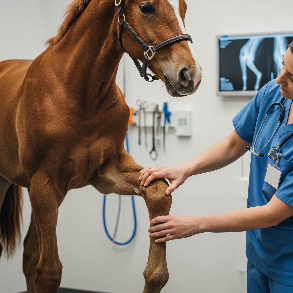
The skeletal system of young horses undergoes dramatic changes during the first several years of life, with bones transitioning from predominantly cartilaginous structures to fully ossified bone. During this critical developmental window, various conditions can arise that compromise the integrity of the musculoskeletal system. These growth-related disturbances, collectively termed developmental orthopedic diseases (DODs), represent a significant concern for horse owners and veterinarians alike.
Developmental orthopedic diseases encompass a spectrum of non-infectious musculoskeletal conditions affecting the growing horse. These disorders stem from disruptions in the normal developmental process of cartilage, bone, and connective tissues, including joint capsules, tendons, and ligaments. Unlike infectious or traumatic conditions, DODs arise from fundamental biological processes going awry during skeletal development, making them distinct challenges in equine medicine.
What Constitutes Developmental Orthopedic Disease?
The term DOD encompasses multiple distinct conditions that, while diverse in presentation and location, share a common thread of disrupted skeletal maturation. These conditions can manifest in various ways throughout the growing horse’s body, affecting different tissues and anatomical locations. Some foals show signs of these conditions from birth, while others develop problems as they age and undergo training.
The conditions classified under DOD include:
- Angular limb deformities (crooked legs)
- Growth plate inflammation (physitis or epiphysitis)
- Subchondral bone cysts (fluid-filled cavities beneath joint cartilage)
- Osteochondrosis and osteochondritis dissecans (cartilage defects)
- Cervical vertebral malformation (spinal canal narrowing)
- Flexural limb deformities (contracted tendons)
The significance of these conditions extends beyond immediate discomfort. Affected horses may experience chronic lameness, reduced athletic potential, and long-term degenerative changes in joints. Early recognition and intervention can substantially influence outcomes and the horse’s future performance capabilities.
Multifactorial Origins: Why Young Horses Develop These Conditions
Developmental orthopedic diseases rarely result from a single cause. Instead, research indicates a complex interplay of multiple factors that collectively contribute to disease development. Understanding these contributing elements is essential for both prevention and management strategies.
Genetic Predisposition
Certain horse breeds demonstrate higher susceptibility to specific developmental orthopedic conditions. Modern horse breeding has emphasized rapid growth and early maturation, traits that paradoxically increase vulnerability to developmental disorders. Thoroughbreds, Quarter Horses, and Warmblood breeds, all selected for accelerated growth potential, frequently experience higher incidence rates of DOD. The heritability of these conditions suggests that breeding decisions directly influence disease prevalence in future generations.
Growth Rate and Metabolic Demands
Foals genetically programmed for rapid growth face unique challenges during skeletal development. When growth plates advance at accelerated rates, the cartilage cells within these structures may not mature appropriately, resulting in defective conversion to bone. This biological mismatch between growth velocity and cellular maturation processes creates conditions favoring skeletal abnormalities. Additionally, obesity resulting from excessive dietary energy compounds these problems by increasing mechanical stress on developing structures.
Nutritional Factors
Nutritional imbalances represent one of the most modifiable risk factors for developmental orthopedic disease. Growing foals and lactating mares require precisely calibrated nutrient profiles to support skeletal development. Deficiencies or imbalances in essential minerals—particularly calcium, phosphorus, copper, and zinc—can disrupt normal bone formation and cartilage maturation. Conversely, overfeeding grain and providing excessive dietary energy accelerates growth beyond what the skeletal system can accommodate, leading to structural failures.
Environmental and Exercise Factors
The physical demands placed on young horses significantly influence skeletal health. Excessive exercise in immature foals can traumatize developing cartilage and growth plates, while insufficient activity fails to provide appropriate stimulus for proper bone remodeling. The balance between adequate exercise and avoiding overuse represents a critical management consideration.
Recognizing the Clinical Presentation
The manifestations of developmental orthopedic disease vary considerably depending on which structures are affected and the severity of the underlying pathology. Horse owners should remain vigilant for early warning signs, as prompt recognition enables timely intervention.
Joint-Related Signs
The most apparent sign of DOD involving joints is excessive fluid accumulation, appearing as noticeable swelling around affected joints. Importantly, swelling associated with osteochondrosis and early developmental disease typically does not present as hot or painful to palpation, distinguishing it from infectious or inflammatory conditions. Joint stiffness often accompanies swelling, and affected horses may demonstrate lameness that varies with exercise intensity and duration. As training begins, latent developmental problems may suddenly become apparent through lameness that was not previously evident.
Growth Plate Abnormalities
When growth plates become inflamed (physitis), the affected areas demonstrate characteristic findings. Joints typically feel warm and firm when palpated and often develop a boxed or squared appearance due to growth plate enlargement. Foals may exhibit visible swelling at the sites where bones meet, particularly around the knees, ankles, and hips. This inflammatory response reflects the underlying disruption of normal ossification processes at the growth plate.
Limb Alignment Issues
Crooked limbs represent another visible manifestation of developmental orthopedic disease. Angular limb deformities present as deviation from normal straight-line alignment when viewing the limb from the front or back. Some foals are born with these deformities, while others develop them progressively during growth. Contracted tendons represent the opposite presentation, where flexor tendons shorten, pulling joints into abnormal flexion positions.
Neurological Manifestations
When developmental orthopedic disease affects the cervical spine, neurological signs predominate. Wobbler syndrome results from malformed vertebrae that narrow the spinal canal, compressing nerve tissue. Affected horses develop incoordination, weakness, and abnormal gait patterns, potentially progressing to severe incapacity.
Diagnostic Approaches and Imaging Strategies
Establishing a definitive diagnosis of developmental orthopedic disease typically requires a combination of clinical evaluation and advanced imaging. Most cases are diagnosed based on characteristic clinical signs combined with radiographic findings.
Clinical Examination
Veterinary assessment begins with a thorough physical examination, including careful observation of limb alignment, gait evaluation, and palpation of joints and growth plates for swelling and sensitivity. In many young horses, lameness may represent the first and only clinical sign, and this lameness frequently does not become apparent until the horse begins training. This delayed presentation underscores the importance of baseline radiographic screening in young horses destined for performance.
Radiographic Evaluation
Radiographs remain the primary diagnostic tool for detecting developmental orthopedic disease. X-ray imaging reveals abnormalities in bone structure, identifies cartilage defects, detects growth plate irregularities, and visualizes subchondral bone cysts. Interestingly, radiographic abnormalities may not correlate perfectly with clinical signs, as some radiographically evident lesions remain asymptomatic while others cause significant problems. Osteochondritis dissecans most frequently affects the hock, stifle, and fetlock joints, locations that should receive particular scrutiny during radiographic evaluation.
Advanced Diagnostic Imaging
When radiographs prove inconclusive or additional detail is needed, advanced imaging modalities such as ultrasound and MRI can provide enhanced visualization of soft tissue structures and early cartilage changes not visible on conventional radiographs.
Timing of Diagnosis and Clinical Implications
A notable characteristic of developmental orthopedic disease is the frequent delay between when lesions actually develop and when they are clinically diagnosed. Osteochondrosis and osteochondritis dissecans typically develop during the first year of life, yet horses often are not diagnosed with these conditions until they are 2-3 years old and enter training. This lag occurs because young foals often show no clinical signs despite having significant radiographic abnormalities, and signs only become apparent when the horse is worked under saddle or in harness.
Management and Treatment Considerations
While developmental orthopedic disease cannot be cured, various management strategies can minimize progression, reduce clinical signs, and optimize the horse’s functional capacity. Treatment approaches are individualized based on the specific condition, severity, and the horse’s intended use.
Nutritional Management
Optimizing nutrition represents one of the most impactful interventions available. This involves ensuring adequate but not excessive caloric intake, providing balanced mineral ratios (particularly calcium and phosphorus), and supplying sufficient trace minerals essential for bone and cartilage formation. Feeding programs should be designed to promote steady, moderate growth rather than rapid weight gain.
Exercise Modification
Appropriate exercise programs support skeletal development without creating excessive mechanical stress. Young horses benefit from regular turnout that allows natural movement patterns, while strenuous training should be delayed until skeletal maturity is achieved.
Monitoring and Early Detection
Regular veterinary examinations throughout the growth period enable early detection of developing problems. Some conditions warrant radiographic screening even in the absence of clinical signs, particularly in high-risk breeds or individuals with affected relatives.
Preventing Developmental Orthopedic Disease
While not all cases can be prevented—particularly those with strong genetic components—implementation of evidence-based management practices reduces incidence and severity:
- Provide balanced nutrition formulated specifically for growing foals and lactating mares
- Avoid overfeeding and limit grain to appropriate levels for age and activity
- Ensure adequate mineral supplementation, especially copper and zinc
- Allow generous turnout for natural movement and exercise
- Delay high-impact exercise and training until skeletal maturity
- Monitor growth rates and body condition regularly
- Consider breeding away from genetic predispositions when possible
Long-Term Prognosis and Performance Implications
The long-term outlook for horses with developmental orthopedic disease varies tremendously based on the specific condition, severity, affected anatomical location, and the intensity of intended use. Some foals with mild angular limb deformities or early physitis may recover completely with appropriate management and grow into sound, capable riding or performance horses. Conversely, severe osteochondritis dissecans affecting critical joints or cervical vertebral malformation may result in chronic lameness or neurological deficits that limit or prevent athletic use.
The presence of developmental orthopedic disease should not automatically condemn a young horse to diminished future potential. Rather, it necessitates individualized assessment, careful management, and realistic expectations about ultimate performance capabilities. Working collaboratively with an equine veterinarian to develop a comprehensive management plan optimizes the horse’s musculoskeletal health and quality of life throughout its lifespan.
References
- Developmental Orthopedic Diseases in Horses — Mad Barn Equine Nutrition. 2024. https://madbarn.com/developmental-orthopedic-diseases-in-horses/
- Developmental Orthopedic Disease in Young, Growing Horses — SmartPak Equine. 2024. https://www.smartpakequine.com/learn-health/developmental-orthopedic-disease
- Developmental Orthopedic Disease (DOD) Causes and Nutritional Solutions — Buckeye Nutrition. 2024. https://www.buckeyenutrition.com/categories/developmental-orthopedic-diseases
- Osteochondrosis: What Horse Owners Should Know — Morris Animal Foundation. 2023. https://www.morrisanimalfoundation.org/article/osteochondrosis-what-horse-owners-should-know
- Developmental Orthopedic Disease in Horses: Part II – Causes and Effects — Vita Flex. 2024. https://www.vitaflex.com/research/developmental-orthopedic-disease
- Developmental Orthopedic Disease in Horses — Kentucky Equine Research. 2024. https://ker.com/equinews/developmental-orthopedic-disease-horses/
- Joint and Bone Abnormalities of the Growing Horse — New England Equine Medical and Surgical Center. 2024. https://www.newenglandequine.com/Articles/Developmental%20Orthopedic%20Disease.pdf



