

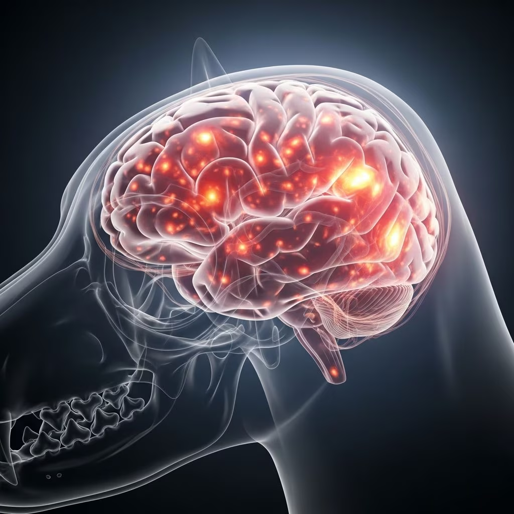
Understanding Meningoencephalitis of Unknown Origin
Meningoencephalitis of unknown origin (MUO) is a serious neurological condition affecting dogs worldwide. This inflammatory disease impacts the brain and the surrounding fluid and tissues, creating significant health challenges for affected pets and their owners. Understanding this condition is essential for recognizing early warning signs and seeking appropriate veterinary care. MUO represents a complex diagnostic puzzle because inflammation is evident through advanced imaging and laboratory analysis, yet the underlying cause remains unidentifiable through standard microscopic examination.
What Exactly is Meningoencephalitis of Unknown Origin?
Meningoencephalitis refers to inflammation of the brain and the cerebrospinal fluid surrounding it, along with the protective tissues encasing the nervous system. In canine patients, meningoencephalitis can be categorized into several distinct types based on characteristic pathological patterns observed during microscopic examination. These include granulomatous meningoencephalomyelitis (GME), necrotizing meningoencephalitis (NME), greyhound meningoencephalitis, and necrotizing leukoencephalitis (NLE).
MUO specifically describes cases where magnetic resonance imaging (MRI) and cerebrospinal fluid (CSF) analysis clearly demonstrate inflammatory, non-infectious central nervous system disease, yet definitive diagnosis through histopathology—microscopic examination of nervous system tissue—is not available. This diagnostic challenge arises when tissue sampling is either impossible, impractical, or when owners decline invasive biopsy procedures. The condition fundamentally represents an autoimmune disorder where the body’s immune system attacks its own nervous system for reasons that remain scientifically unclear.
Breed Predisposition and Risk Factors
MUO shows distinct patterns in its occurrence across different dog populations, with certain breeds demonstrating significantly higher susceptibility than others. Understanding these risk factors helps veterinarians identify at-risk dogs and enables preventative discussions with dog owners.
Breed Susceptibility
Small-breed dogs are disproportionately affected by MUO, suggesting a strong genetic component to the disease. Research has specifically identified genetic risk factors in the Pug and Maltese breeds, making these populations particularly vulnerable. Other small breeds including Chihuahuas and toy breeds show increased prevalence. While large-breed dogs can occasionally develop the disease, they represent a minority of MUO cases, reinforcing the breed-specific pattern observed in this condition.
Age and Gender Patterns
Demographic analysis reveals that females are affected more frequently than males, representing an interesting sex-based predisposition. Affected dogs are typically over six months of age, though the disease can present at various life stages. Young to middle-aged small-breed females represent the highest-risk demographic for developing this condition.
Clinical Presentation and Symptoms
The clinical signs of MUO vary considerably depending on which specific brain regions experience inflammation. Recognizing these neurological manifestations is crucial for prompt veterinary evaluation and diagnosis.
Primary Neurological Signs
Dogs with MUO commonly present with several characteristic neurological symptoms. Seizures represent one of the most common clinical manifestations, ranging from localized seizures affecting specific body areas to generalized seizures involving the entire body. Muscle tremors occur frequently and may be progressive or intermittent. Vision loss or complete blindness can develop as inflammation affects visual processing areas of the brain.
Vestibular signs such as dizziness, loss of balance, and falling over indicate involvement of the brain’s balance centers. Head tilting is a classic neurological sign associated with MUO and vestibular disease. These signs create obvious behavioral changes that alert owners to their dog’s declining neurological status.
Behavioral and Movement Changes
Beyond primary neurological signs, affected dogs may display abnormal behaviors including compulsive circling, which suggests disorientation and altered consciousness. Some dogs appear to experience pain or discomfort, potentially indicating meningeal inflammation causing pain signals. Partial or complete paralysis can develop in severe cases, significantly impacting the dog’s mobility and quality of life.
Disease Progression Timeline
MUO symptoms may develop suddenly in some cases, creating acute onset of neurological crisis. In other affected dogs, signs progress slowly over weeks to months, creating a more insidious presentation that may initially be overlooked or attributed to aging. This variable presentation timeline makes accurate diagnosis challenging and underscores the importance of comprehensive neurological evaluation whenever neurological signs appear in dogs.
Diagnostic Approach and Procedures
Diagnosing MUO requires a systematic approach combining multiple diagnostic modalities to rule out other conditions and confirm inflammatory CNS disease.
Initial Evaluation
Veterinary evaluation begins with a thorough physical examination and baseline blood work assessing complete blood cell counts and internal organ function. This initial screening identifies systemic abnormalities that might explain neurological signs through non-neurological causes such as metabolic disease, organ dysfunction, or systemic infection.
When initial blood work appears normal and no obvious systemic causes explain the neurological abnormalities, veterinarians typically recommend infectious disease testing. This comprehensive testing rules out infectious causes of CNS inflammation including bacterial infections, viral infections like distemper, and parasitic infections that could produce similar neurological signs.
Specialized Neurological Testing
Dogs requiring definitive diagnosis of potential MUO are typically referred to veterinary neurologists for advanced diagnostic procedures. Two critical tests aid in MUO diagnosis: magnetic resonance imaging (MRI) and cerebrospinal fluid (CSF) analysis.
Magnetic Resonance Imaging
MRI uses powerful magnetic fields to generate detailed three-dimensional images of internal brain structures. This advanced imaging modality allows neurologists to assess brain anatomy and identify characteristic abnormalities suggesting meningoencephalitis. The procedure requires general anesthesia because dogs must remain completely still for extended periods—typically 30 to 60 minutes—to obtain high-quality diagnostic images. MRI can reveal abnormal signal patterns, swelling, or structural changes consistent with inflammatory brain disease.
Cerebrospinal Fluid Analysis
During the anesthesia period required for MRI, neurologists perform a cerebrospinal fluid tap—a specialized procedure collecting a small fluid sample from around the spinal cord. The brain and spinal cord are surrounded by cerebrospinal fluid, which acts as a shock absorber and protective cushion for the nervous system. In a CSF tap, a long needle is carefully positioned to withdraw a small amount of this fluid for laboratory analysis.
Laboratory examination of CSF includes assessment of fluid composition and microscopic evaluation to identify cell types present. Inflammatory meningoencephalitis produces characteristic CSF changes including elevated protein levels and specific white blood cell patterns. These results directly assess for inflammatory disease of the nervous system.
Treatment and Management Strategies
MUO is typically caused by underlying autoimmune dysfunction where the immune system inappropriately attacks nervous system tissue. Treatment therefore focuses on immune system suppression to reduce inflammatory damage to the brain.
Immunosuppressant Medications
Dogs diagnosed with or suspected of having MUO are treated with immunosuppressant drugs that reduce immune system activity and decrease CNS inflammation. Several medications are commonly employed in MUO management:
Corticosteroids including prednisone and dexamethasone represent first-line therapy, working rapidly to suppress immune system activation and reduce inflammation. These medications are often used as initial treatment and sometimes as monotherapy in milder cases.
Cyclosporine (Atopica) is a specialized immunosuppressant that targets specific immune cells, often used in combination with corticosteroids or as an alternative when corticosteroids prove insufficient or produce unacceptable side effects.
Azathioprine (Imuran) provides additional immunosuppressive capability and is frequently used as part of multi-drug immunosuppressive protocols for more severe or refractory cases.
Treatment Protocols
Most MUO cases require combination immunosuppressive therapy using multiple medications at varying doses to achieve adequate disease control while minimizing medication side effects. Treatment protocols are individualized based on disease severity, response to initial therapy, and medication tolerance. Long-term management often requires gradual dose reduction over months to find the minimum effective medication doses maintaining disease control.
Frequently Asked Questions About Meningoencephalitis of Unknown Origin
Q: Is MUO contagious to other dogs?
A: No, MUO is not contagious. This autoimmune condition develops due to genetic predisposition and immune system dysfunction in individual dogs, not from infectious agents that could spread between animals.
Q: Can MUO be prevented in at-risk breeds?
A: Currently, no proven prevention exists for MUO. While genetic predisposition is established in certain breeds, owners cannot prevent disease development. Early recognition of symptoms remains crucial for initiating treatment promptly.
Q: What is the prognosis for dogs with MUO?
A: Prognosis varies significantly depending on disease severity, brain regions affected, treatment response, and individual dog factors. Some dogs achieve remission with appropriate treatment, while others experience chronic disease requiring ongoing management.
Q: How long does MUO treatment typically last?
A: Treatment duration varies considerably. Some dogs require lifelong immunosuppressive therapy, while others achieve remission and gradually discontinue medications. Treatment plans are individualized based on disease response and clinical progression.
Q: Are there any dietary modifications beneficial for MUO dogs?
A: While no specific diet treats MUO, maintaining overall health through appropriate nutrition supports immune function and general wellness. Discuss dietary modifications with your veterinarian based on your dog’s individual needs and medication requirements.
Q: Can I breed dogs with MUO?
A: Given the identified genetic predisposition to MUO in certain breeds, responsible breeders should avoid breeding affected dogs. Breeding decisions should consider the established genetic risk factors in susceptible breeds.
Living with a Dog Diagnosed with MUO
Dogs diagnosed with MUO require consistent veterinary monitoring and medication management. Regular follow-up appointments allow veterinarians to assess treatment effectiveness, monitor for medication side effects, and adjust therapy as needed. Owners should maintain detailed records of symptom changes and medication responses to facilitate optimal care adjustments. Many dogs with MUO achieve good quality of life with appropriate treatment, though managing a chronic neurological condition requires commitment and ongoing veterinary collaboration.
References
- Meningoencephalitis of Unknown Origin – VCA Animal Hospitals — VCA Inc. 2024. https://vcahospitals.com/know-your-pet/meningoencephalitis-of-unknown-origin
- Of potential new treatment targets and polythetic approach in meningoencephalitis of unknown origin — Frontiers in Veterinary Science. 2024. https://www.frontiersin.org/journals/veterinary-science/articles/10.3389/fvets.2024.1465689/full
- Meningoencephalitis of Unknown Aetiology (MUA) — Fitzpatrick Referrals. 2024. https://www.fitzpatrickreferrals.co.uk/neurology/meningoencephalitis-of-unknown-aetiology-mua/
- Prognosis in meningoencephalitis of unknown origin in dogs: Risk factors and outcome — Journal of Veterinary Internal Medicine. 2024. https://onlinelibrary.wiley.com/doi/10.1111/jvim.17037



