

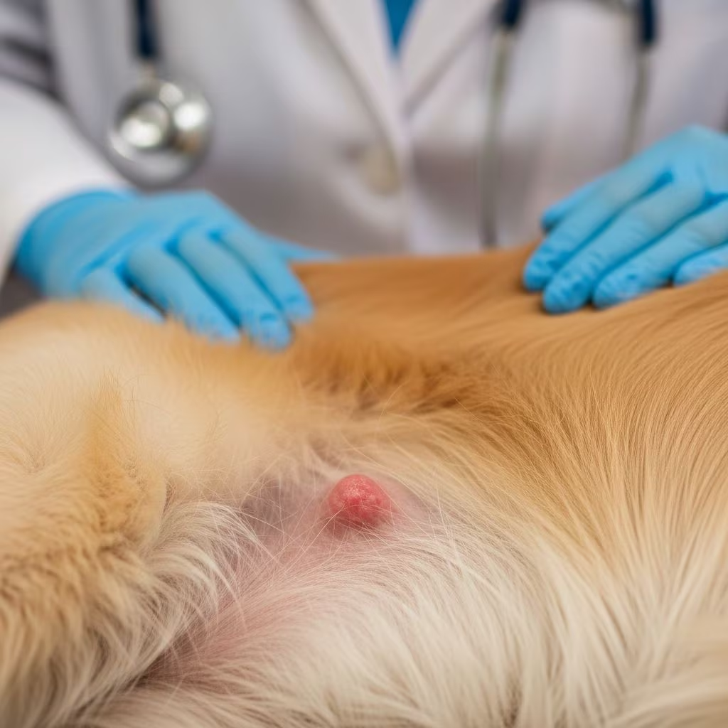
Understanding Mast Cell Tumors in Dogs
Mast cell tumors (MCTs) represent one of the most common skin cancers found in dogs, accounting for a significant portion of cutaneous neoplasms in the canine population. These tumors develop from mast cells, which are immune cells located throughout the body, particularly in the skin and gastrointestinal tract. Understanding the nature of these tumors, their progression, and available treatment options is essential for pet owners facing this diagnosis. The unpredictable behavior of mast cell tumors means that each case requires careful evaluation and individualized treatment planning by your veterinarian.
What Are Mast Cells and Why Do They Matter?
Mast cells are immune cells that play a vital role in the body’s inflammatory and allergic responses. These cells contain granules filled with various chemical mediators, including histamine, serotonin, and heparin. When mast cells become cancerous, they can proliferate uncontrollably and form tumors. One of the most challenging aspects of mast cell tumors is their tendency to degranulate, releasing large quantities of histamine into the bloodstream. This degranulation can trigger a cascade of clinical signs ranging from local skin reactions to systemic complications affecting multiple organ systems.
Clinical Signs and Symptoms
The presentation of mast cell tumors varies considerably depending on the tumor’s location, grade, and disease burden. Many dogs with early-stage disease may show no clinical signs beyond the visible tumor mass itself. However, as the disease progresses, owners may observe several concerning symptoms:
– Scratching, biting, or licking at a skin mass- Vomiting and diarrhea- Bloody stools- Lethargy and decreased activity levels- Loss of appetite- Edema or swelling around the tumor site- Wheal formation (raised, inflamed areas on the skin)
More severe systemic signs typically develop in dogs with larger disease burden or internal spread of the cancer. In extreme cases, massive degranulation can lead to anaphylaxis, a life-threatening allergic reaction. Dogs with metastatic disease—where cancer has spread to lymph nodes, liver, or spleen—are more likely to experience gastrointestinal complications, including severe vomiting and diarrhea.
Diagnosis and Staging
Accurate diagnosis and staging of mast cell tumors is critical for determining appropriate treatment and predicting outcomes. When a skin mass is suspected to be a mast cell tumor, your veterinarian will begin with a cytological examination. This involves taking a small sample of cells from the tumor, typically using fine needle aspiration, and examining them under a microscope. This initial assessment helps confirm the diagnosis and provides the first indication of tumor grade.
Grading System
Mast cell tumors are classified into three grades based on their microscopic appearance and behavior:
– Grade I (Low-Grade): Well-differentiated tumors with slow growth rates and excellent prognosis- Grade II (Intermediate-Grade): Moderately differentiated tumors with variable behavior- Grade III (High-Grade): Poorly differentiated, aggressive tumors with rapid growth and poor prognosis
Low-grade tumors have significantly better outcomes, with many dogs achieving complete remission through surgical removal alone. High-grade tumors are more challenging to treat and have higher rates of recurrence and metastasis.
Staging Investigations
Beyond grading, your veterinarian will perform staging investigations to determine if the cancer has spread. These may include:
– Physical examination of regional lymph nodes- Fine needle aspiration of nearby lymph nodes- Abdominal ultrasound to evaluate internal organs- Blood work and biochemical profiles- Evaluation of sentinel lymph nodes for metastatic involvement
The presence of lymph node involvement significantly impacts prognosis. Patients with lymph node metastasis have a 5-fold increased risk of tumor progression and a 4-fold increased risk of tumor-related death compared to those without lymph node involvement.
Treatment Options
The treatment approach for mast cell tumors depends on multiple factors including the tumor grade, stage, size, location, number of tumors, and the dog’s overall health status. Several treatment modalities are available, often used in combination for optimal outcomes.
Surgical Removal
Surgical excision remains the preferred and most effective treatment for mast cell tumors, particularly for low-grade localized disease. The surgical approach involves removing the tumor with adequate margins of healthy tissue surrounding it. For low-grade tumors, surgical removal often provides excellent long-term control and may result in complete cure. Most stage I cases are resolved through surgical intervention alone.
The extent of surgical margins depends on the tumor grade and location. Your surgeon will work to remove the tumor while preserving as much healthy tissue as possible. Complete surgical removal provides the best opportunity for long-term disease control in lower-grade tumors with no evidence of spread.
Chemotherapy
Chemotherapy becomes particularly important in cases where the tumor cannot be completely removed surgically, has spread to multiple organs, or is high-grade. Several chemotherapy options are available:
Tyrosine Kinase Inhibitors (TKIs)
Toceranib phosphate (Palladia) is currently the only tyrosine kinase inhibitor labeled specifically for canine mast cell tumor treatment in the United States. These medications work by inhibiting cellular signaling pathways that promote tumor cell growth. TKIs are typically administered orally at home, making them convenient for long-term management. Overall response rates for TKIs range from 30% to 60% with variable progression-free intervals. Costs generally range from $400 to $700 per month depending on the dog’s weight. TKIs can be combined with radiation therapy or traditional chemotherapy in cases of gross or microscopic disease, though this combination may increase the risk of adverse events.
Intralesional Therapy
Intralesional treatments are injected directly into the tumor and include options such as triamcinolone, a long-acting steroid. In clinical studies, triamcinolone demonstrated an overall response rate of 67% (complete and partial responses combined). When combined with oral corticosteroids, the median time to progression extended to 114 days compared to 28 days with triamcinolone alone.
Tigilanol tiglate injection represents an exciting newer option in intralesional therapy. Approximately 75% of patients with cytologically low-grade tumors achieved complete response after a single treatment, increasing to nearly 90% with a second treatment. Even for high-grade tumors, response rates reached 44% after one treatment and 67% after two treatments. A significant advantage of tigilanol tiglate is that most responders have significantly smaller wounds than the 2- to 3-centimeter margins previously recommended with traditional surgery.
Immunotherapy
Gilvetmab is an innovative immunotherapy administered as an intravenous infusion every two weeks for up to ten treatments. This treatment showed an overall response rate of 46% in canine patients with mast cell tumors. As with many immunotherapies, patients require monitoring for pseudoprogression, a phenomenon where tumor size temporarily increases due to resurgent inflammation. This local growth can be misinterpreted as treatment failure and may lead to premature discontinuation of therapy. Pseudoprogression can delay visible tumor shrinkage by 8 to 12 weeks, making patience and careful monitoring essential during early treatment phases.
Corticosteroid Therapy
Prednisone and other systemic corticosteroids have demonstrated inhibitory effects on mast cell viability and proliferation. These medications also effectively decrease clinical signs associated with histamine degranulation, including edema, itching, and pain. While single-agent corticosteroid therapy is economical and accessible, responses are often short-lived, lasting only a few weeks. However, when used as neoadjuvant therapy (before surgery), corticosteroids can achieve maximal response within 7 to 14 days, during which surgical excision should be scheduled. Corticosteroids can also serve as adjuvant therapy (after surgery) or sole treatment in palliative care situations, with monthly costs typically ranging from $30 to $250.
Radiation Therapy
Radiation therapy becomes valuable when mast cell tumors cannot be completely removed through surgery due to their location or size. This treatment involves multiple visits to a specialty hospital where the dog is anesthetized and radiation is applied directly to the tumor. Radiation therapy may also be combined with surgical removal for intermediate or high-grade tumors to improve local control and reduce recurrence rates.
Supportive Care and Management
Dogs with mast cell tumors require comprehensive supportive care to manage symptoms related to histamine degranulation and maintain quality of life. Many veterinarians prescribe histamine antagonists to decrease signs associated with mast cell degranulation and elevated plasma histamine concentrations.
Histamine Management
Two types of histamine antagonists serve different purposes:
– H1 Antagonists (such as diphenhydramine): Primarily limit cutaneous inflammation and allergic reactions- H2 Antagonists (such as famotidine): Critically important as approximately 30% of dogs with mast cell tumors show evidence of gastrointestinal ulceration at necropsy
For dogs with high disease burden—such as those with large, weeping tumors or disseminated metastasis—proton pump inhibitors like omeprazole may provide more targeted blockade and superior long-term efficacy compared to standard H2 antagonists.
Managing Treatment Side Effects
Depending on the treatment modality chosen, dogs may experience side effects requiring additional management. Common side effects include nausea, vomiting, lethargy, and decreased appetite. Medications to address these issues include:
– Entyce (capromorelin): An appetite stimulant administered daily to help maintain food intake, energy levels, and body weight- Cerenia (maropitant): An anti-nausea medication effective for managing vomiting and nausea that often improves appetite
Recovery After Surgery
Dogs recovering from surgical removal of low-grade mast cell tumors typically require two weeks of restricted activity and rest. Recovery management includes pain medications, antihistamines such as Benadryl, and use of a recovery collar to prevent licking or trauma to the surgical site. After two weeks, sutures are usually removed and normal activity can gradually resume, though your veterinarian will provide specific guidance based on your individual dog’s recovery progress.
Prognosis and Long-Term Outcomes
The prognosis for mast cell tumors varies dramatically based on multiple prognostic factors. The good news is that most mast cell tumors are low to intermediate grade, with high-grade aggressive tumors being much less common. Low- and intermediate-grade tumors are less likely to grow back after complete surgical removal, and many dogs achieve excellent long-term survival.
For low-grade tumors with no evidence of spread, surgery provides the best long-term control. A majority of stage I cases can be considered cured through surgical removal. In contrast, high-grade masses that have spread aggressively to lymph nodes and internal organs require multimodal treatment and carry a guarded prognosis.
Close monitoring is extremely important, especially for dogs with a history of mast cell tumors, as these dogs may be prone to developing new tumors (not metastatic recurrence but new primary tumors). Regular veterinary checkups help detect any new masses early when treatment options are most effective.
When to Consider Palliative Care
For some dogs with advanced or metastatic mast cell tumors, the focus may shift from curative treatment to palliative care aimed at maintaining quality of life. Considerations for palliative care or euthanasia may include:
– Uncontrollable weight loss, vomiting, and loss of appetite despite appetite stimulants and nausea medications- Recurrent severe gastrointestinal signs from tumor degranulation causing sudden onset bloody vomiting and diarrhea with severe dehydration requiring hospitalization- Inability to maintain comfort despite available pain management options- Significant decline in quality of life and overall functioning
Frequently Asked Questions
Q: Is there a cure for mast cell tumors in dogs?
A: If the mast cell tumor has a low grade and low mitotic index, surgical removal may result in a complete cure. However, close monitoring is important as dogs with a history of mast cell tumors may develop new tumors. High-grade tumors typically have poorer outcomes despite treatment.
Q: How do I know if my dog’s mast cell tumor has spread?
A: Your veterinarian can determine if the tumor has spread through lymph node evaluation, abdominal ultrasound, and blood work. The most common sites of metastasis are regional lymph nodes, liver, and spleen. Lymph node involvement significantly worsens the prognosis.
Q: What is the success rate of surgery for mast cell tumors?
A: For low-grade tumors with no evidence of spread, surgical removal is highly successful and often curative. Most stage I cases are resolved through surgery alone. Success rates decrease for high-grade and metastatic tumors.
Q: Are there any new treatments for mast cell tumors?
A: Yes, newer treatment options include tigilanol tiglate injection (an intralesional therapy with high response rates), gilvetmab (an immunotherapy), and tyrosine kinase inhibitors like toceranib phosphate. Your veterinarian can discuss which newer options may benefit your dog.
Q: How long can a dog live with a mast cell tumor?
A: Survival time depends on the tumor grade, stage, and whether it has metastasized. Dogs with completely removed low-grade tumors may live several years or longer. High-grade or metastatic tumors have shorter survival times, though treatment can extend life and maintain quality.
Q: What causes mast cell tumors in dogs?
A: The exact cause of mast cell tumors is not fully understood. There may be genetic predisposition in certain breeds, and environmental factors likely play a role, but no single cause has been definitively identified.
References
- Simplifying the Approach to Canine Mast Cell Tumors — Today’s Veterinary Practice. 2024. https://todaysveterinarypractice.com/oncology/simplifying-the-approach-to-canine-mast-cell-tumors/
- Mast Cell Tumor (Mastocytoma) in Dogs — PetMD. 2024. https://www.petmd.com/dog/conditions/cancer/mast-cell-tumor-in-dogs
- Mast Cell Tumors in Dogs — VCA Animal Hospitals. 2024. https://vcahospitals.com/know-your-pet/mast-cell-tumors-in-dogs
- Diagnosis, Prognosis and Treatment of Canine Cutaneous and Subcutaneous Mast Cell Tumors — National Center for Biotechnology Information (NCBI/PubMed Central). 2023. https://pmc.ncbi.nlm.nih.gov/articles/PMC8870669/
- Medical Oncology: Mast Cell Tumors in Dogs — North Carolina State University College of Veterinary Medicine. 2024. https://hospital.cvm.ncsu.edu/services/small-animals/cancer-oncology/oncology/mast-cell-tumors/
- Mast Cell Tumors (Mastocytomas) in Dogs — Small Door Veterinary. 2024. https://www.smalldoorvet.com/learning-center/medical/mast-cell-tumors-mastocytomas
- Mast Cell Tumors — American College of Veterinary Surgeons. 2024. https://www.acvs.org/small-animal/mast-cell-tumors/



