

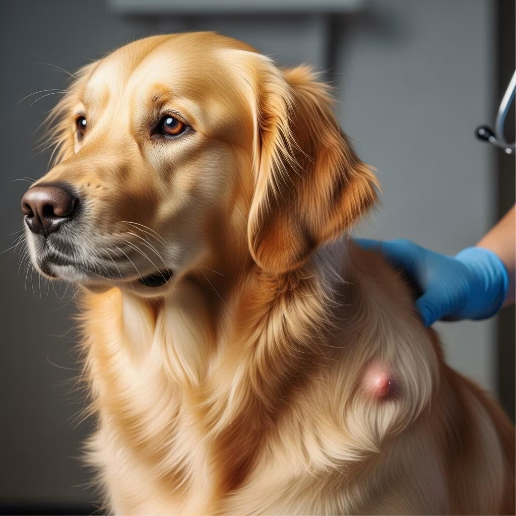
Mast cell tumors represent one of the most prevalent skin cancers affecting dogs, often appearing as lumps that vary in severity from benign to highly aggressive. These tumors arise from mast cells, which play a key role in allergic responses and immune function, but can lead to serious health issues when they proliferate uncontrollably.
Understanding the Biology of Canine Mast Cell Tumors
Mast cells are part of the immune system, releasing chemicals like histamine during allergic reactions. In dogs, when these cells undergo cancerous transformation, they form mast cell tumors (MCTs), primarily in the skin but potentially spreading to lymph nodes, liver, spleen, or other organs. Breeds such as Boxers, Bulldogs, and Retrievers show higher susceptibility, though any dog can develop them.
The tumors’ unpredictable nature stems from their ability to degranulate, releasing histamine and other mediators that cause local swelling, redness, or systemic effects like gastrointestinal upset.
Recognizing Symptoms and Early Warning Signs
Early detection improves outcomes significantly. Common signs include:
- A firm, raised lump on the skin that may change size, often reddish or ulcerated.
- Itching, scratching, or licking at the site.
- Swelling around the tumor due to histamine release.
- Systemic issues like vomiting, diarrhea, lethargy, or loss of appetite in advanced cases.
- Rarely, anaphylactic shock with collapse.
Tumors can mimic benign cysts or insect bites, underscoring the need for veterinary evaluation of any new skin mass.
Diagnostic Approaches for Accurate Assessment
Veterinarians start with a physical exam and fine-needle aspiration (FNA) to examine cells under a microscope, providing a preliminary grade. For confirmation, a full biopsy with histopathology is essential, assessing mitotic index and other features.
Staging involves checking regional lymph nodes via cytology or sentinel lymph node mapping, plus imaging like ultrasound or X-rays for internal spread. Bloodwork monitors for anemia or organ dysfunction caused by degranulation products.
| Diagnostic Tool | Purpose | Key Insights |
|---|---|---|
| Fine-Needle Aspiration | Initial cell sampling | Quick, detects 70-90% of MCTs |
| Biopsy & Histopathology | Definitive grading | Patnaik or Kiupel grading system |
| Lymph Node Evaluation | Metastasis check | Cytology or biopsy for staging |
| Imaging (Ultrasound/CT) | Systemic spread | Liver, spleen assessment |
Grading and Staging: Predicting Tumor Behavior
Grading classifies tumors as low (grade I/II, good prognosis), high (grade III, aggressive), using systems like Patnaik (three-tier) or Kiupel (two-tier). High mitotic index (>5 per high-power field) signals poor outcomes.
Staging from I (localized skin) to IV (distant metastasis) guides therapy. Prognostic panels analyze KIT mutations, aiding TKI therapy decisions.
Primary Treatment: Surgical Intervention
Surgery remains the cornerstone for most MCTs, aiming for wide margins (2-3 cm) and one fascial plane deep. Clean margins yield cure rates over 90% for low-grade tumors.
For incomplete excisions, re-operation or adjunct therapies follow. Multiple tumors may require staged surgeries.
Radiation Therapy Options
MCTs respond well to radiation, especially post-incomplete surgery. Fractionated protocols (15-18 sessions, 46-54 Gy total) achieve 75-96% local control at 3-5 years.
Stereotactic radiation offers fewer sessions for select cases, minimizing side effects like skin ulceration.
Systemic Therapies for Advanced Cases
For metastatic or high-risk MCTs, chemotherapy protocols like vinblastine/prednisone (weekly then spaced doses) target microscopic disease.
Tyrosine kinase inhibitors (TKIs) such as toceranib (Palladia) or masitinib excel against KIT-mutated tumors, shrinking bulky masses and extending survival.
Emerging options include water-soluble paclitaxel, showing 30% response rates in non-resectable grade II/III tumors.
Innovative Local Therapies: Stelfonta and Beyond
Stelfonta (tigilanol tiglate) is an FDA-approved injectable for non-metastatic MCTs up to 5 cm. It disrupts tumor membranes, cuts blood supply, and boosts immunity, destroying 87% of tumors in 1-2 injections with dogs resuming normal activity in 14 days.
Electrochemotherapy and intralesional agents provide alternatives for challenging locations.
Supportive Care to Manage Degranulation
Antihistamines (diphenhydramine/Benadryl) prevent wheal formation and GI issues. H2 blockers (famotidine) and proton pump inhibitors (omeprazole) protect the stomach. Prednisone offers rapid symptom relief and occasional remission, though short-lived.
- Administer Benadryl 1-2 mg/lb every 8-12 hours.
- Monitor for vomiting; add gastroprotectants.
- Avoid NSAIDs due to ulcer risk.
Prognosis Factors and Survival Expectations
Low-grade, clean-margin tumors boast >95% 2-year survival. High-grade or metastatic cases drop to 6-12 months without aggressive therapy, improved by TKIs to 1-2 years.
Lymph node involvement halves survival; early intervention is critical.
| Factor | Good Prognosis | Poor Prognosis |
|---|---|---|
| Grade | I/II | III |
| Margins | Clean | Incomplete |
| Location | Trunk/limbs | Mucocutaneous |
| Metastasis | None | Lymph nodes/organs |
Living with a Dog Diagnosed with MCTs
Post-treatment monitoring includes monthly exams initially, then quarterly, with owner vigilance for new lumps. Holistic supports like omega-3s and antioxidants may aid, but evidence is anecdotal.
Quality of life remains high for most; euthanasia decisions hinge on systemic spread and symptoms.
FAQs on Mast Cell Tumors in Dogs
What breeds are most at risk? Boxers, Boston Terriers, Labrador Retrievers, and Pugs have genetic predispositions.
Can MCTs be prevented? No sure prevention, but spaying/neutering may reduce risk in predisposed breeds. Regular skin checks aid early detection.
Is chemotherapy painful for dogs? Side effects are mild (vomiting, lethargy) in 20-30% of cases, managed with supportive care.
How quickly does Stelfonta work? Tumor necrosis begins within days, full resolution in 4-6 weeks.
What if surgery isn’t possible? Radiation, TKIs, or Stelfonta offer viable alternatives with good local control.
Advances in Research and Future Directions
Ongoing trials explore immunotherapy and novel TKIs. KIT mutation testing refines personalized medicine, promising better outcomes for high-risk cases.
References
- Simplifying the Approach to Canine Mast Cell Tumors — Today’s Veterinary Practice. 2023. https://todaysveterinarypractice.com/oncology/simplifying-the-approach-to-canine-mast-cell-tumors/
- Canine mast cell tumors: diagnosis, treatment, and prognosis — PMC (NCBI). 2020-07-01. https://pmc.ncbi.nlm.nih.gov/articles/PMC7337164/
- About STELFONTA — Stelfonta Official Site. 2023. https://stelfonta.com/about-stelfonta/
- FAQs: Mast Cell Tumours in Dogs — The Pet Oncologist. 2023. https://www.thepetoncologist.com/faqs-mast-cell-tumours-in-dogs
- Mast Cell Tumors in Dogs — NC State Veterinary Hospital. 2023. https://hospital.cvm.ncsu.edu/services/small-animals/cancer-oncology/oncology/mast-cell-tumors/



