

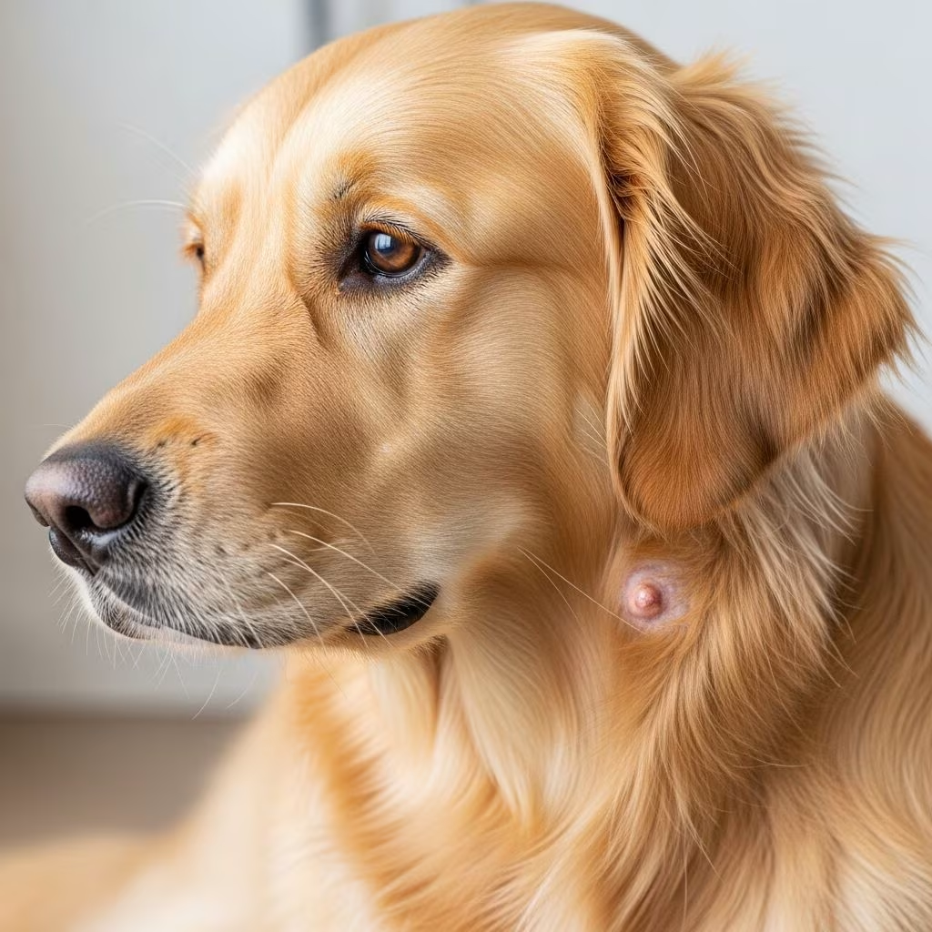
Mast cell tumors represent one of the most frequently diagnosed skin cancers in dogs, accounting for a significant portion of canine malignancies. These abnormal growths develop from mast cells, which are immune system components responsible for releasing histamine and other inflammatory mediators. When mast cells become cancerous and multiply uncontrollably, they form tumors that can affect not only the skin but potentially spread to internal organs. Understanding this condition is essential for dog owners because early detection and appropriate intervention can dramatically improve survival rates and quality of life.
What Are Mast Cell Tumors and Why Do They Form?
Mast cells normally function as part of the immune system’s defense mechanism, helping dogs respond to allergens and irritants. However, when genetic mutations occur within these cells, they can transform into cancerous growths. The exact triggers that cause this transformation remain incompletely understood, though researchers have identified certain breeds as having higher predisposition rates. Mast cell tumors develop most commonly on the skin, particularly in areas prone to friction or trauma, but they can also emerge in the digestive tract, spleen, liver, and bone marrow in more advanced cases.
These tumors vary significantly in their biological behavior. Some mast cell tumors grow slowly and remain localized, while others display aggressive characteristics with rapid growth and metastatic potential. This variability in tumor behavior directly influences treatment decisions and long-term prognosis, making accurate grading and staging crucial for determining the optimal management approach.
Recognizing the Early Signs and Getting Diagnosed
Dog owners often notice mast cell tumors as unexplained lumps or bumps on their pet’s skin. These growths may appear anywhere on the body but commonly develop on the legs, trunk, or genital area. Some tumors itch persistently, causing dogs to lick or scratch excessively, while others remain asymptomatic until discovered during routine veterinary examinations.
When internal organs become affected, dogs may display systemic symptoms including vomiting, diarrhea, loss of appetite, lethargy, or difficulty breathing. These signs result from mast cell degranulation—the release of histamine and other inflammatory mediators that disrupt normal body functions. Diagnosis typically begins with a physical examination followed by cytology or biopsy to confirm the presence of mast cells and determine the tumor’s grade, a critical factor in treatment planning.
The Grading System and What It Means for Your Dog
Veterinary pathologists classify mast cell tumors using a grading system that predicts biological behavior and guides treatment decisions. Low-grade tumors (grades I and II) grow slowly and rarely metastasize, offering favorable prognoses when treated appropriately. High-grade tumors (grade III) exhibit aggressive characteristics, spread more readily to distant sites, and require more intensive treatment protocols.
The grading system also considers mitotic index—the frequency of cell division—and anatomical location. Tumors in certain locations such as the perineum, inguinal area, or digits tend to behave more aggressively than those in other body regions. Understanding your dog’s specific tumor grade allows your veterinary oncologist to develop a personalized treatment strategy tailored to the tumor’s biological potential.
Surgical Intervention: The Foundation of Treatment
Surgical removal represents the primary treatment modality for most mast cell tumors and offers the best opportunity for cure in many cases. The surgical goal extends beyond simply removing the visible tumor; it requires excising the entire lesion with adequate margins of healthy tissue surrounding it. Surgical margins typically range from 5 millimeters to 40 millimeters, depending on tumor characteristics and location.
For low-grade tumors with adequate surgical margins and no evidence of metastasis, surgical removal alone frequently results in complete remission. Dogs with low-grade tumors often achieve excellent long-term survival following appropriate surgery. Following surgical removal, tissue samples are sent for pathological examination to confirm complete excision and reassess tumor grade, which may influence recommendations for additional therapy.
Recovery from surgical removal typically requires approximately two weeks of restricted activity, pain management, and antihistamine administration. Pet owners must closely monitor surgical sites for signs of infection or complications and maintain follow-up appointments as scheduled.
Chemotherapy Protocols for Aggressive Tumors
Dogs with high-grade tumors or evidence of metastasis benefit from systemic therapy to eliminate cancer cells throughout the body. Chemotherapy becomes particularly important when surgical margins prove incomplete or when tumors are not surgically resectable.
Common chemotherapy protocols utilize vinblastine combined with prednisone, administered in carefully timed intervals. An eight-dose protocol typically treats dogs with aggressive tumors without gross measurable disease. When tumors remain palpable after surgery, treatment adjusts based on tumor response, continuing weekly doses initially, then transitioning to every-two-week intervals, and eventually spacing treatments as far apart as possible while maintaining tumor control.
Tyrosine kinase inhibitors (TKIs) such as toceranib phosphate (Palladia) and masitinib represent oral medications given at home, offering convenience compared to injectable chemotherapy. These targeted drugs specifically inhibit mechanisms that allow mast cells to survive and multiply. Studies demonstrate that toceranib phosphate achieves response rates of approximately 37% in dogs with bulky, non-resectable grade II or III tumors. Emerging research explores combining toceranib phosphate with traditional chemotherapy, achieving response rates around 71%.
Radiation Therapy for Challenging Locations
Radiation therapy proves invaluable when mast cell tumors develop in locations unsuitable for surgery or when surgical excision leaves microscopic disease behind. Mast cell tumors demonstrate remarkable radiosensitivity, responding well to fractionated radiation protocols. Treatment typically involves 15 to 18 daily or every-other-day sessions, delivering total doses between 46 and 54 gray. Success rates are impressive, with 75% to 96% of dogs achieving complete local remission with no evidence of regrowth at 3 to 5 years post-radiation.
Radiation therapy sessions require anesthesia and specialized equipment available only at veterinary specialty facilities, necessitating travel and multiple visits. Despite these practical considerations, radiation offers an excellent treatment option for dogs whose tumors cannot be completely removed surgically.
Emerging Therapies and Innovative Approaches
Veterinary oncology continues advancing with new therapeutic options. STELFONTA represents an injectable treatment that works through three mechanisms: disrupting tumor cell membranes, cutting off blood supply, and stimulating the immune system to fight cancer. This treatment destroys 87% of mast cell tumors with just one or two injections, allowing dogs to return to normal activities within 14 days. However, not all dogs or tumors qualify as suitable candidates for this therapy.
Supportive and Palliative Care Strategies
Beyond tumor-directed treatments, supportive medications manage symptoms resulting from mast cell degranulation and maintain quality of life. Antihistamines such as diphenhydramine (Benadryl) reduce histamine effects, while antacids including omeprazole or famotidine protect the gastrointestinal tract from acid damage. Corticosteroids like prednisolone provide rapid anti-inflammatory effects, sometimes producing complete remission within one to two days of administration, though remission duration typically averages one to two months.
For dogs experiencing metastatic disease with internal tumor spread, appetite loss and gastrointestinal distress become significant concerns. Medications such as capromorelin (Entyce) stimulate appetite and help maintain body weight, while maropitant (Cerenia) controls nausea and vomiting. These supportive treatments significantly improve comfort and maintain nutritional status during treatment courses.
Treatment Cost Considerations
Financial planning becomes important when developing comprehensive treatment strategies. A comparison of treatment expenses demonstrates the range of options available:
| Treatment Modality | Typical Cost Range | Administration Method |
|---|---|---|
| Steroids (Palliative) | $30 – $250 monthly | Oral medication daily |
| Tyrosine Kinase Inhibitors | $400 – $700 monthly | Oral every other day or three times weekly |
| Chemotherapy | Variable; typically several hundred dollars per treatment | Injectable, administered in veterinary clinic |
| Radiation Therapy | Variable; substantial cost for full protocol | Multiple anesthetized sessions over weeks |
Discussing costs openly with your veterinary team helps identify realistic options aligned with your circumstances and ensures your dog receives appropriate care.
Understanding Prognosis and Long-Term Outcomes
Prognosis varies dramatically based on tumor characteristics and selected treatment. Dogs with completely excised low-grade tumors often experience excellent long-term survival, potentially living many years after treatment. The median survival time for dogs with inguinal or perineal mast cell tumors treated aggressively reaches approximately 37 months, demonstrating the potential for extended survival with appropriate intervention.
High-grade tumors or tumors with metastatic spread present more challenging prognoses, though multimodal treatment approaches—combining surgery, chemotherapy, and radiation—significantly improve outcomes compared to single-treatment modalities. Individual factors including overall health, age, immune function, and response to initial therapy all influence long-term survival expectations.
Frequently Asked Questions
- How quickly do mast cell tumors grow? Growth rates vary significantly. Low-grade tumors typically progress slowly over months or years, while high-grade tumors may expand rapidly within weeks.
- Can mast cell tumors be prevented? No definitive prevention exists, though maintaining healthy weight, minimizing skin trauma, and regular veterinary screenings enable early detection when prognosis remains most favorable.
- What happens if we don’t treat mast cell tumors? Untreated tumors continue expanding and may eventually metastasize to vital organs, causing systemic disease and reduced survival times.
- Are certain dog breeds more susceptible? Yes, specific breeds including Boxers, Pugs, Boston Terriers, and Bulldogs demonstrate higher predisposition, though any breed can develop mast cell tumors.
- Can my dog undergo multiple treatments? Absolutely. Multimodal treatment combining surgery, chemotherapy, and radiation often provides superior outcomes compared to single-treatment approaches.
Moving Forward with Your Dog’s Care
A diagnosis of mast cell tumor understandably concerns dog owners, yet significant advances in veterinary oncology provide multiple effective treatment options. Working closely with your veterinarian and potentially consulting with a board-certified veterinary oncologist ensures your dog receives the most appropriate care based on individual tumor characteristics and overall health status. Regular monitoring, compliance with recommended treatments, and attention to supportive care maximize your dog’s comfort and survival potential, allowing many dogs to continue enjoying quality life for extended periods following diagnosis and treatment.
References
- Mast Cell Tumors in Dogs — Paws at Peace. Accessed January 2026. https://pawsatpeace.com/mast-cell-tumors-in-dogs/
- Canine Mast Cell Tumors: Diagnosis, Treatment, and Prognosis — National Center for Biotechnology Information/PubMed Central. 2020. https://pmc.ncbi.nlm.nih.gov/articles/PMC7337164/
- About STELFONTA: How It Works to Treat Canine Mast Cell Tumors — STELFONTA Official. Accessed January 2026. https://stelfonta.com/about-stelfonta/
- Mast Cell Tumor — PWD Foundation. Accessed January 2026. https://www.pwdfoundation.org/health/cancer/mast-cell-tumor/
- Mast Cell Tumours in Dogs — The Pet Oncologist. Accessed January 2026. https://www.thepetoncologist.com/faqs-mast-cell-tumours-in-dogs
- Medical Oncology: Mast Cell Tumors in Dogs — North Carolina State University College of Veterinary Medicine. Accessed January 2026. https://hospital.cvm.ncsu.edu/services/small-animals/cancer-oncology/oncology/mast-cell-tumors/
- Mast Cell Tumors in Dogs — VCA Animal Hospitals. Accessed January 2026. https://vcahospitals.com/know-your-pet/mast-cell-tumors-in-dogs



