

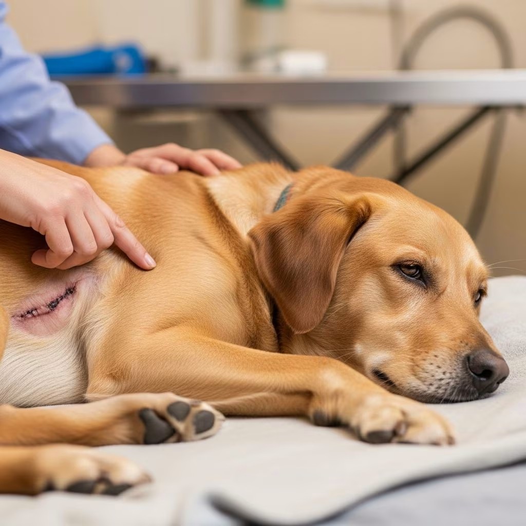
Seromas represent a frequent yet typically benign occurrence in the postoperative phase for dogs, manifesting as fluid-filled swellings near surgical sites. These collections of serous fluid arise when the body responds to tissue disruption by producing plasma-like liquid in areas lacking proper support, often resolving naturally within weeks under vigilant care.
Understanding the Nature of Seromas
A seroma forms due to the accumulation of clear, watery fluid—primarily serum from blood plasma combined with inflammatory cells—in subcutaneous spaces created during surgery. This happens because surgical removal of tissue or tumors leaves “dead space” that the body fills as part of the healing process, exacerbated by the dog’s natural movements or reactions to sutures. Unlike abscesses, which involve pus and infection, seromas are sterile unless complicated.
Surgical seromas commonly appear above incision lines, particularly in high-motion areas like the ventral midline (belly center), where approximately 10% of spayed dogs develop them. Non-surgical seromas can occur elsewhere from trauma but are less relevant here. They feel soft, fluctuant (fluid-filled), and non-painful initially, distinguishing them from firm tumors or inflamed hematomas (blood collections).
Common Surgical Procedures Linked to Seroma Development
Certain operations heighten seroma risk due to extensive tissue manipulation or location over active joints. Key examples include:
- Spay surgeries: Prevalent in puppies, whose high energy makes confinement challenging, leading to fluid buildup in abdominal areas.
- Tumor or mass removals: Especially large lumps, where significant dissection creates voids.
- TPLO (Tibial Plateau Leveling Osteotomy): Knee surgeries for cruciate ligament tears, where mobility promotes fluid accumulation.
- Other soft tissue procedures: Such as hernia repairs or exploratory surgeries in the ventral midline.
These procedures involve subcutaneous layers, where fluid easily pools if tissues are overly dissected or handled roughly.
Recognizing Seroma Symptoms in Your Dog
Early detection aids effective management. Look for a soft, squishy lump under the skin near the incision, often appearing 3-10 days post-op. It may grow with activity but feels cool or neutral, not hot. Normal signs include:
- Fluctuant texture when gently palpated.
- Translucent or clear if fluid leaks (rare).
- No associated pain unless large.
Warning signs requiring immediate vet attention:
| Concerning Symptom | Why It’s Serious |
|---|---|
| Pain, heat, redness | Possible infection or abscess formation |
| Fever, lethargy, appetite loss | Systemic infection risk |
| Rapid growth (>2 inches) or leaking pus | May need drainage to prevent rupture |
| Hardening or persistence beyond 3 weeks | Capsule formation blocking absorption |
Distinguish from normal swelling (peaks at 3-5 days, then recedes) or reactions to sutures. Always consult your vet for confirmation via palpation or ultrasound if unsure.
Home Care Strategies for Mild Seromas
Most small seromas (under 2 inches, non-painful) self-resolve in 2-3 weeks as the body reabsorbs fluid. Support this with conservative measures:
- Activity restriction: Crate rest or leash walks only for 10-14 days to minimize motion-induced fluid production. Sedation may help energetic pups.
- Warm compresses: Apply for 5-10 minutes, 3-4 times daily to enhance circulation and absorption. Use a clean cloth soaked in warm (not hot) water.
- Cold compresses initially: First 48 hours post-op to reduce inflammation, then switch to warm.
- E-collar use: Prevent licking or scratching, which introduces bacteria.
- Massage gently: If vet-approved, light stroking disperses fluid without puncturing.
Critical rule: Never squeeze, drain, or puncture at home—this risks severe infection.
Professional Veterinary Interventions
When home care fails or complications arise, vets employ tiered treatments based on size and symptoms:
- Aspiration: Sterile needle and syringe remove fluid in-clinic. Repeat if reaccumulates, though fluid often refills quickly.
- Drain placement: For persistent cases, a closed suction or Penrose drain allows ongoing fluid exit over days.
- Medications: Antibiotics for infection, analgesics for pain, corticosteroids to curb inflammation.
- Surgical revision: Rare last resort for encapsulated seromas, involving debridement and tighter closure.
Studies on closed suction drains in canine wounds show they reduce seroma rates by managing fluid production relative to body weight, informing modern protocols.
Prevention Tips for Surgical Seromas
Proactive steps minimize risk:
- Choose experienced surgeons who minimize tissue trauma and use proper closure techniques.
- Request drains for high-risk procedures like large mass excisions.
- Enforce strict rest post-op; consider short-term sedatives for active dogs.
- Monitor incisions daily and report issues early.
For spays or TPLOs, pre-op discussions about activity management are key.
Expected Recovery Timeline
| Time Post-Op | Typical Progression |
|---|---|
| Days 1-3 | Initial swelling peaks; start cold compresses. |
| Weeks 1-2 | Seroma may appear; warm compresses, rest critical. |
| Weeks 2-3 | Fluid absorption; size decreases if uncomplicated. |
| Beyond 3 weeks | Vet evaluation if persistent. |
Frequently Asked Questions (FAQs)
Will my dog’s seroma go away on its own?
Yes, most do within 2-3 weeks with rest and compresses, but monitor closely.
Is a seroma dangerous for my dog?
Usually benign, but infection risks make vet check-ups essential if symptomatic.
How can I tell if it’s infected?
Look for heat, redness, pain, discharge, or behavioral changes like lethargy.
Should I drain the seroma myself?
No—risks infection; always seek professional aspiration.
Can seromas recur after treatment?
Possible if activity isn’t restricted; drains reduce recurrence.
Long-Term Outlook and When to Follow Up
With proper management, dogs recover fully without scarring or mobility issues. Schedule a 10-14 day recheck post-op, regardless. Seromas rarely impact surgery benefits like spays improving health or TPLOs restoring joint function. Persistent cases may need imaging to rule out deeper issues.
Empower your dog’s healing by staying informed and proactive. Early intervention ensures minor setbacks don’t derail recovery.
References
- What is the management of seroma formation post-operatively in a dog? — DrOracle.ai. 2024. https://www.droracle.ai/articles/131712/what-is-the-management-of-seroma-formation-post-operatively-in
- Seromas in Dogs: Causes, Pictures, Treatment & Prevention — PetHelpful. 2023. https://pethelpful.com/dogs/seromas-bump-on-a-dogs-surgery-incision
- What Is This Lump at My Pet’s Incision Site? — Spay Neuter Vets. 2024. https://www.spayneutervets.com/blog/post-surgery-lump-seroma
- What is A Seroma In Dogs And How To Prevent It — Kingsdale Animal Hospital. 2023. https://www.kingsdale.com/what-is-a-seroma-in-dogs-and-how-to-prevent-it
- Detecting and Treating Seroma in Dogs — Petcube. 2024. https://petcube.com/blog/seroma-in-dogs/
- Preventing & Treating Seroma After Cancer Surgery in Pets — VetFolio. 2023. https://www.vetfolio.com/courses/seroma-prevention-and-managemet-to-drain-or-not-to-drain



