

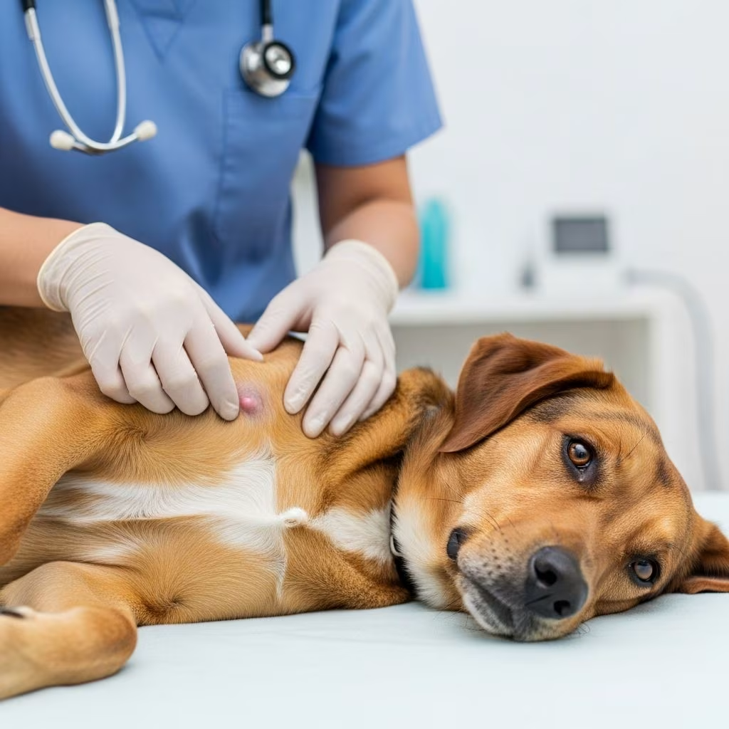
Mammary tumors represent one of the most prevalent forms of cancer affecting female dogs, often manifesting as firm lumps along the abdominal chain of glands. Early identification through routine checks can significantly enhance prognosis, as many such growths respond well to intervention when detected promptly.
Understanding the Anatomy of Canine Mammary Glands
Dogs possess a unique arrangement of mammary tissue, featuring five pairs of glands stretching from the chest to the groin area, totaling ten nipples. These structures serve lactation purposes but are prone to neoplastic changes, particularly in unspayed females where hormonal influences play a key role. Tumors typically emerge in the posterior glands nearer the hind legs, though any gland can be involved.
Male dogs experience these tumors far less frequently due to minimal mammary tissue development, but when they occur, outcomes tend to be more aggressive. Hormones like estrogen and progesterone stimulate cellular proliferation in these glands, elevating risk over multiple heat cycles.
Risk Factors Driving Mammary Tumor Development
Several elements contribute to the onset of mammary tumors. Intact females face the highest susceptibility, with risk escalating alongside the number of heat cycles endured. Spaying prior to the first heat slashes incidence by over 99%, while post-first heat intervention reduces it by about 75%, and after second heat by roughly 25%.
- Age: Incidence peaks between 7-11 years.
- Breed predispositions: Breeds like Poodles, Spaniels, and Dachshunds show higher rates.
- Hormonal history: Prolonged exposure from pregnancies or false heats amplifies danger.
- Obesity: Excess fat may promote estrogen production.
Environmental factors, including chemical exposures, warrant further study but do not override hormonal dominance.
Recognizing Early Warning Signs
Initial detection often happens during casual petting or grooming when owners notice irregularities. Common indicators include:
- Firm, nodular lumps beneath the skin near nipples, varying from pea-sized to golf ball dimensions.
- Discharge from nipples, ranging from clear to bloody or pus-like.
- Swelling, redness, or ulceration over affected areas.
- Enlarged nearby lymph nodes in armpits or groin.
As tumors advance and potentially metastasize, systemic effects emerge:
| Early Local Signs | Advanced Systemic Signs |
|---|---|
| Lumps in mammary chain | Weight loss |
| Nipple discharge | Lethargy and weakness |
| Skin inflammation/ulceration | Appetite reduction |
| Pain on palpation | Coughing or breathing issues |
These manifestations mimic benign conditions like mastitis or cysts, underscoring veterinary evaluation necessity.
Step-by-Step Home Examination Techniques
Monthly self-checks empower owners to spot anomalies early. Position your dog comfortably on their back during a relaxed belly rub session.
- Visual scan: Inspect the underbelly for asymmetry, swelling, or discoloration from chest to groin.
- Gentle palpation: Use fingertips to probe each nipple area, feeling for hard, fixed masses versus soft, movable ones.
- Check lymph nodes: Feel under front legs and near hindquarters for swelling.
- Monitor behavior: Note licking, discomfort, or appetite shifts.
Perform this post-bath when skin is clean. Any new lump merits immediate vet consultation.
Diagnostic Approaches in Veterinary Practice
Vets initiate with physical exams, assessing lump characteristics: size, firmness, mobility, and ulceration. History review covers spay status, heat cycles, and medications.
Further tests include:
- Fine-needle aspiration or biopsy: Cells extracted for cytological or histopathological analysis to distinguish benign from malignant.
- Imaging: Chest X-rays detect lung metastases; abdominal ultrasound evaluates organ spread.
- Blood panels: Complete blood count, chemistry, and urinalysis gauge surgical candidacy.
- Lymph node sampling: Checks for regional invasion.
Staging classifies tumors from I (small, localized) to IV (distant spread), guiding therapy. Approximately 50% of tumors prove malignant, with 50% of those metastasizing, commonly to lungs or lymph nodes.
Treatment Modalities for Optimal Outcomes
Management hinges on tumor stage, patient health, and owner preferences. Surgery remains cornerstone.
Surgical Interventions:
- Lumpectomy: Removes isolated masses.
- Mastectomy: Excises single or chain of glands; simple for one, radical for extensive.
Post-op pathology determines adjunct needs. Chemotherapy suits inflammatory or metastatic cases, using drugs like doxorubicin. Radiation targets inoperable tumors. Median survival post-surgery exceeds 1-2 years for benign, 6-12 months for malignant without spread.
Preventive Strategies Centered on Spaying
Elective ovariohysterectomy before puberty nearly eliminates risk. For intact older dogs, weigh surgery benefits against mammary findings. Obesity control and routine exams bolster prevention.
Prognosis Factors Influencing Longevity
Benign tumors boast excellent prognosis post-removal. Malignant ones vary: small, non-metastasized yield 80-90% one-year survival; metastatic drop to under 20%. Histological grade, surgical margins, and lymph involvement predict success.
FAQs on Mammary Tumors in Dogs
Can male dogs get mammary tumors?
Yes, though rare, affecting about 1% of cases, often more aggressive.
Is every lump cancerous?
No, half are benign; biopsy confirms.
How often should I check my dog?
Monthly, plus annual vet exams.
Does spaying cure existing tumors?
No, but halts progression in hormone-sensitive types.
What breeds are most at risk?
Poodles, English Setters, Terriers, and Spaniels.
Supporting Your Dog Through Diagnosis and Recovery
Post-treatment, monitor for recurrence via check-ups. Nutrition rich in antioxidants aids healing. Emotional support through gentle play sustains quality of life.
Owners play pivotal roles: vigilance saves lives. Collaborate with vets and oncologists for tailored plans.
References
- Breast cancer in dogs — Blue Cross. 2023. https://www.bluecross.org.uk/advice/dog/health-and-injuries/breast-cancer-in-dogs
- Breast Cancer in Dogs — Shoreland Animal Hospital. 2024-05-15. https://www.shorelandanimalhospital.com/site/blog/2024/05/15/breast-cancer-dogs
- Breast Cancer in Dogs — Atlantic Coast Veterinary. 2022-01-15. https://www.atlanticcoastvet.com/site/blog-long-island-vet/2022/01/15/breast-cancer-dog
- Breast Cancer in Dogs — PetCure Oncology. 2023. https://petcureoncology.com/breast-cancer-in-dogs/
- 12 Subtle Signs of Cancer in Dogs — PetMD. 2024. https://www.petmd.com/dog/conditions/signs-of-cancer-in-dogs
- Medical Oncology: Canine Mammary Tumors — NC State Veterinary Hospital. 2024. https://hospital.cvm.ncsu.edu/services/small-animals/cancer-oncology/oncology/canine-mammary-tumors/
- Malignant Mammary Tumors in Dogs — VCA Animal Hospitals. 2024. https://vcahospitals.com/know-your-pet/mammary-tumors-in-dogs-malignant



