

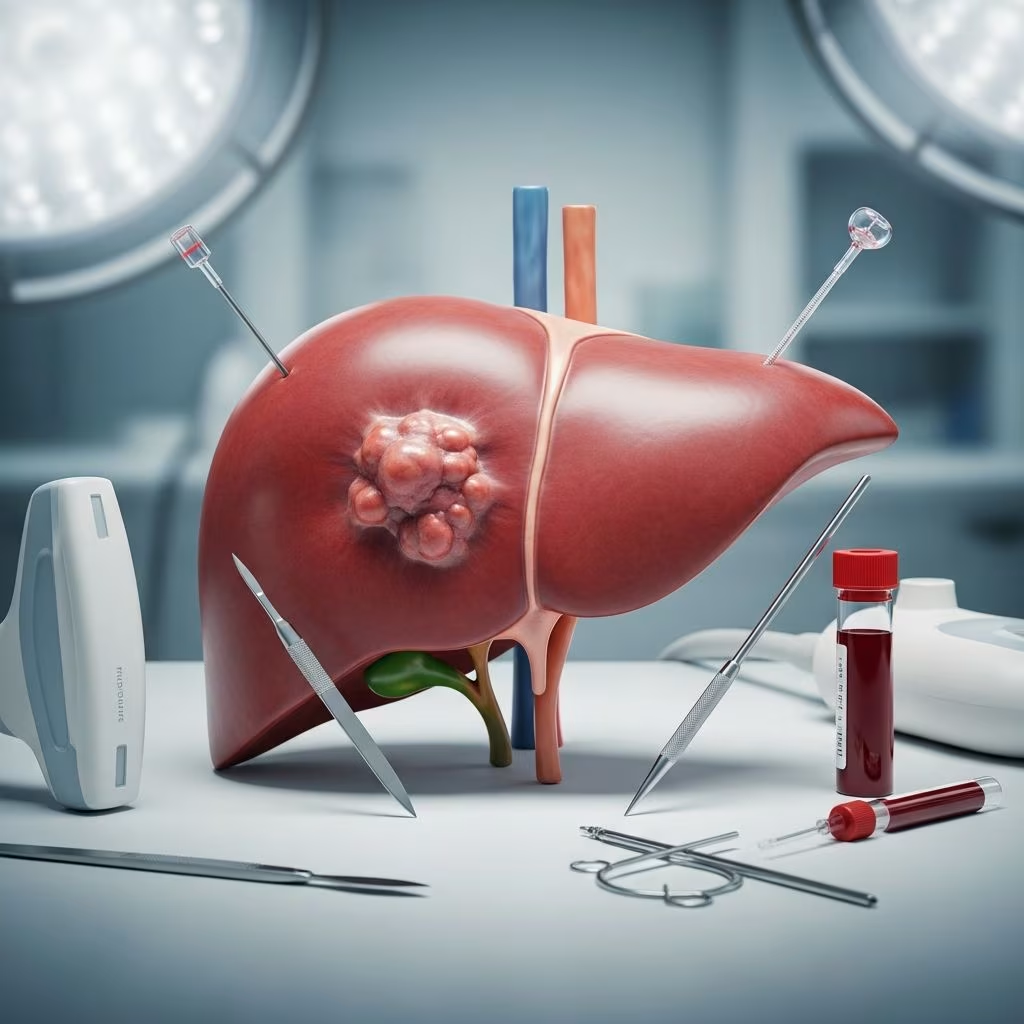
Understanding Hepatic Neoplasia in Equine and Ruminant Populations
Hepatic neoplasia represents an uncommon but significant health challenge in large animal veterinary medicine. While primary liver tumors occur with relatively low frequency in horses and ruminants, their presence carries substantial clinical implications for affected animals. The spectrum of neoplastic conditions affecting the liver in these species encompasses several distinct pathological entities, each with unique characteristics regarding presentation, progression, and prognosis.
The comparative rarity of primary hepatic malignancies in large animals contrasts sharply with their occurrence in other species, yet understanding their biology remains essential for practitioners managing complex cases. Unlike in companion animals where hepatocellular carcinoma dominates the neoplastic landscape, large animal hepatic tumors present with greater morphological diversity and frequently involve secondary neoplastic invasion from systemic diseases.
Classification of Primary Hepatic Malignancies in Large Animals
Primary liver tumors in horses and ruminants fall into several distinct histopathological categories. The most commonly encountered form is hepatocellular carcinoma, which originates directly from the hepatic parenchyma. Additionally, cholangiocarcinoma represents a significant neoplastic entity arising from the biliary epithelium. Lymphoma, though typically recognized as a systemic hematopoietic malignancy, frequently manifests hepatic involvement. Hepatoblastoma, a rare embryonal tumor, has been documented in equine populations.
Secondary hepatic involvement occurs when neoplastic processes originating elsewhere disseminate to liver tissue. Metastatic melanoma, mesothelioma, and carcinomas originating from gastric, renal, or mammary tissues represent common sources of secondary hepatic involvement. The distinction between primary and secondary neoplasia carries substantial diagnostic and prognostic significance.
Systemic Lymphosarcoma and Hepatic Manifestation
Lymphosarcoma emerges as the predominant hematopoietic malignancy in equine populations, with hepatic involvement occurring in a substantial proportion of affected animals. Research indicates that approximately 41% of horses diagnosed with lymphosarcoma demonstrate neoplastic liver involvement, while splenic involvement occurs in as many as 37% of cases. This widespread infiltration reflects the systemic nature of lymphoid malignancies and their predilection for lymphoid tissues throughout the body.
The hepatic manifestations of lymphosarcoma have been documented across multiple ruminant species, including cattle, llamas, alpacas, and goats. The metastatic nature of lymphoid malignancies necessitates comprehensive staging protocols to assess the extent of disease burden throughout affected animals.
Clinical Presentation and Observable Signs
Animals affected by primary hepatic carcinoma typically present with a constellation of clinical signs reflecting progressive hepatic dysfunction. Lethargy and unexplained weight loss represent the most prominent initial observations by animal caretakers. As disease progresses, additional manifestations emerge, including progressive abdominal enlargement, erythrocytosis, persistent hypoglycemia, icterus, and ultimately hepatic failure.
The clinical course of cholangiocarcinoma demonstrates a characteristic pattern, with pronounced weight loss occurring prominently before the development of frank hepatic failure. This temporal relationship provides clinicians with a diagnostic window during which intervention may be considered, though by the time clinical signs manifest, significant disease burden typically exists.
Hepatoblastoma, when present, presents with distinctive systemic manifestations including erythrocytosis and large areas of extramedullary hematopoiesis, reflecting the tumor’s unique biological behavior. Metastasis to the thoracic cavity has been documented in equine cases, extending the potential systemic burden of disease.
Laboratory and Biochemical Findings
Hepatic enzyme elevations provide crucial diagnostic clues in animals with liver malignancy. Hepatocellular and biliary enzyme concentrations increase in response to hepatic carcinoma or cholangiocarcinoma, reflecting both direct tumor-related hepatocellular damage and cholestasis. Gamma-glutamyl transferase activity demonstrates particularly pronounced elevation in affected horses, often reaching very high levels that suggest biliary obstruction or cholestasis.
Serum alpha-fetoprotein concentration elevation has been identified as a potential marker in cases of hepatoblastoma or hepatocellular carcinoma. This oncofetal antigen, normally present at minimal levels in adult animals, may be upregulated by certain neoplastic hepatic tissues, providing an adjunctive diagnostic tool.
Cytological examination of peritoneal fluid and peripheral blood may reveal atypical lymphocytes or lymphoblasts in animals with hepatic lymphoid involvement, supporting diagnosis of systemic lymphoproliferative disease.
Diagnostic Imaging and Ultrasonographic Assessment
Ultrasonographic examination represents the primary imaging modality for detecting splenic and hepatic neoplasia in large animal populations. This non-invasive technique provides real-time visualization of hepatic architecture and facilitates identification of focal or diffuse pathology. Hepatocellular carcinomas demonstrate characteristic uniform appearance on ultrasonographic examination, facilitating differentiation from other hepatic pathologies.
The acoustic properties of neoplastic tissue often differ substantially from normal hepatic parenchyma, allowing experienced ultrasonographers to identify suspicious lesions. However, ultrasonographic findings must be corroborated with additional diagnostic testing to establish definitive diagnosis and exclude benign processes.
Advanced imaging modalities such as computed tomography may be utilized in select cases to assess tumor extent and staging, particularly when considering surgical management or evaluating for distant metastasis.
Tissue Diagnosis and Histopathological Confirmation
Liver biopsy with subsequent microscopic examination represents the gold standard for definitive diagnosis of hepatic neoplasia. The presence and character of hepatic neoplasia can be confirmed through this invasive diagnostic procedure, which provides definitive histopathological classification.
Multiple biopsy techniques exist, including percutaneous needle approaches, laparoscopic methods, and open surgical biopsy. The selection of approach depends upon clinician preference, available equipment, and patient factors. Biopsy procedures generally carry acceptable risk profiles in large animals when performed by experienced practitioners, though hemorrhagic complications remain a potential concern given the highly vascular nature of hepatic tissue.
Clinical Manifestations in Ruminant Populations
Ruminant animals with hepatic neoplasia frequently present with clinical signs dominated by tumor growth in non-hepatic locations. The multifocal nature of neoplastic processes in ruminants means that clinical signs often reflect involvement of lymph nodes, abomasum, cardiac structures, reproductive organs, or spinal cord rather than purely hepatic dysfunction.
This presentation pattern reflects the systemic nature of many ruminant malignancies, particularly lymphoid neoplasias, which show strong predilection for involvement of multiple organ systems simultaneously. Consequently, diagnostic approaches in ruminants must encompass comprehensive staging to identify all sites of neoplastic involvement.
Prognostic Considerations and Disease Trajectory
The prognosis for large animals diagnosed with primary hepatic neoplasia remains guarded to poor. The progressive nature of hepatic malignancy, combined with the relatively advanced disease burden typically present at the time of diagnosis, limits therapeutic options and long-term survival prospects. Early recognition, though challenging given the non-specific initial clinical signs, offers the best opportunity for intervention.
The extent of neoplastic spread at diagnosis carries profound prognostic significance. Animals with localized, resectable lesions may have better outcomes than those with extensive infiltration or documented metastatic disease. However, the rarity of these tumors means that large series documenting long-term outcomes remain limited in the veterinary literature.
Treatment Approaches and Therapeutic Considerations
Surgical resection represents the most definitive therapeutic option for hepatic neoplasia when tumors are focal, non-metastasized, and located within resectable hepatic regions. The remarkable regenerative capacity of hepatic tissue permits removal of substantial liver mass while maintaining adequate hepatic function. The surgical approach must carefully weigh the extent of tissue removal against the need to preserve sufficient functional liver parenchyma.
For malignant tumors that are extensive or have metastasized, systemic chemotherapy may be considered for palliative purposes, though efficacy remains limited. The unique challenge of hepatic tumors lies in the organ’s capacity to metabolically inactivate chemotherapeutic agents, reducing drug efficacy. Additionally, the poor blood supply to tumor centers limits chemotherapy drug penetration.
Supportive care remains an essential component of management for all affected animals. Nutritional support, management of secondary hepatic dysfunction, and alleviation of clinical signs contribute to quality of life during the disease course.
Distinguishing Primary from Secondary Hepatic Involvement
| Feature | Primary Hepatic Neoplasia | Secondary Hepatic Involvement |
|---|---|---|
| Origin | Arises directly from hepatic tissue | Originates elsewhere, metastasizes to liver |
| Prevalence | Uncommon | More frequent than primary |
| Examples | Hepatocellular carcinoma, cholangiocarcinoma | Melanoma, mesothelioma, gastric carcinoma |
| Diagnostic Focus | Liver biopsy, imaging | Primary tumor identification, staging |
| Treatment Approach | Surgical resection when possible | Depends on primary tumor type and extent |
Frequently Asked Questions About Hepatic Neoplasia in Large Animals
Can large animals survive hepatic neoplasia without treatment?
Survival without treatment is possible but limited. The progressive nature of hepatic malignancy means disease will advance, leading to increasing hepatic dysfunction and eventual hepatic failure. The duration of untreated disease varies based on tumor type and extent.
How is hepatic neoplasia definitively diagnosed?
Liver biopsy with histopathological examination provides definitive diagnosis. While ultrasonography raises suspicion and guides biopsy site selection, tissue examination is required for conclusive classification and characterization of tumor type.
What percentage of large animals with hepatic disease have neoplasia?
Primary hepatic neoplasia remains uncommon in horses and ruminants. While exact prevalence statistics are limited in veterinary literature, neoplasia represents a small percentage of diagnosed hepatic disease in these populations.
Are there breed or age predispositions for hepatic tumors in large animals?
The rarity of primary hepatic neoplasia in large animals limits comprehensive epidemiological data regarding breed or age predispositions. Further research is needed to establish whether specific populations carry elevated risk.
Summary and Clinical Implications
Hepatic neoplasia in large animals represents a challenging diagnostic and therapeutic problem for veterinary practitioners. The relative rarity of these conditions, combined with non-specific clinical presentations, often results in delayed diagnosis. However, awareness of the potential for primary and secondary hepatic malignancy enables practitioners to maintain appropriate clinical suspicion and pursue diagnostic confirmation through imaging and tissue examination.
Recognition of the clinical signs associated with hepatic malignancy—particularly unexplained lethargy, progressive weight loss, and abdominal distension—should prompt thorough hepatic evaluation. Laboratory abnormalities including elevated hepatic enzymes provide additional diagnostic clues warranting further investigation. Ultrasonographic examination facilitates identification of focal hepatic lesions, while liver biopsy confirms diagnosis and guides prognostic and therapeutic decision-making.
Though prognosis for animals with hepatic neoplasia remains guarded, surgical intervention may be considered in select cases with localized, resectable disease. The regenerative capacity of hepatic tissue provides hope for animals undergoing partial hepatectomy, though long-term survival data remain limited. A comprehensive, evidence-based approach to diagnosis and management offers the best opportunity to provide meaningful care for large animals affected by this serious condition.
References
- Management of Hepatic Neoplasia in Humans and Dogs — National Institutes of Health, National Center for Biotechnology Information. 2022. https://pmc.ncbi.nlm.nih.gov/articles/PMC9505178/
- Hepatic Neoplasia in Large Animals — Merck Veterinary Manual. 2024. https://www.msdvetmanual.com/digestive-system/hepatic-disease-in-large-animals/hepatic-neoplasia-in-large-animals
- Morphological Classification of Neoplastic Disorders of the Canine Liver — Comparative Hepatology Research Group. 2021. https://comparativehepatology.org/wp-content/uploads/2021/09/chapter_9.pdf
- Information for Oncology Clients: Liver Tumors in Dogs and Cats — University of Pennsylvania School of Veterinary Medicine. 2025. https://www.vet.upenn.edu/wp-content/uploads/2025/05/liver-tumors.pdf
- Canine Hepatobiliary Tumors — North Carolina State University College of Veterinary Medicine. 2024. https://hospital.cvm.ncsu.edu/services/small-animals/cancer-oncology/oncology/medical-oncology-canine-hepatobiliary-tumors/



