

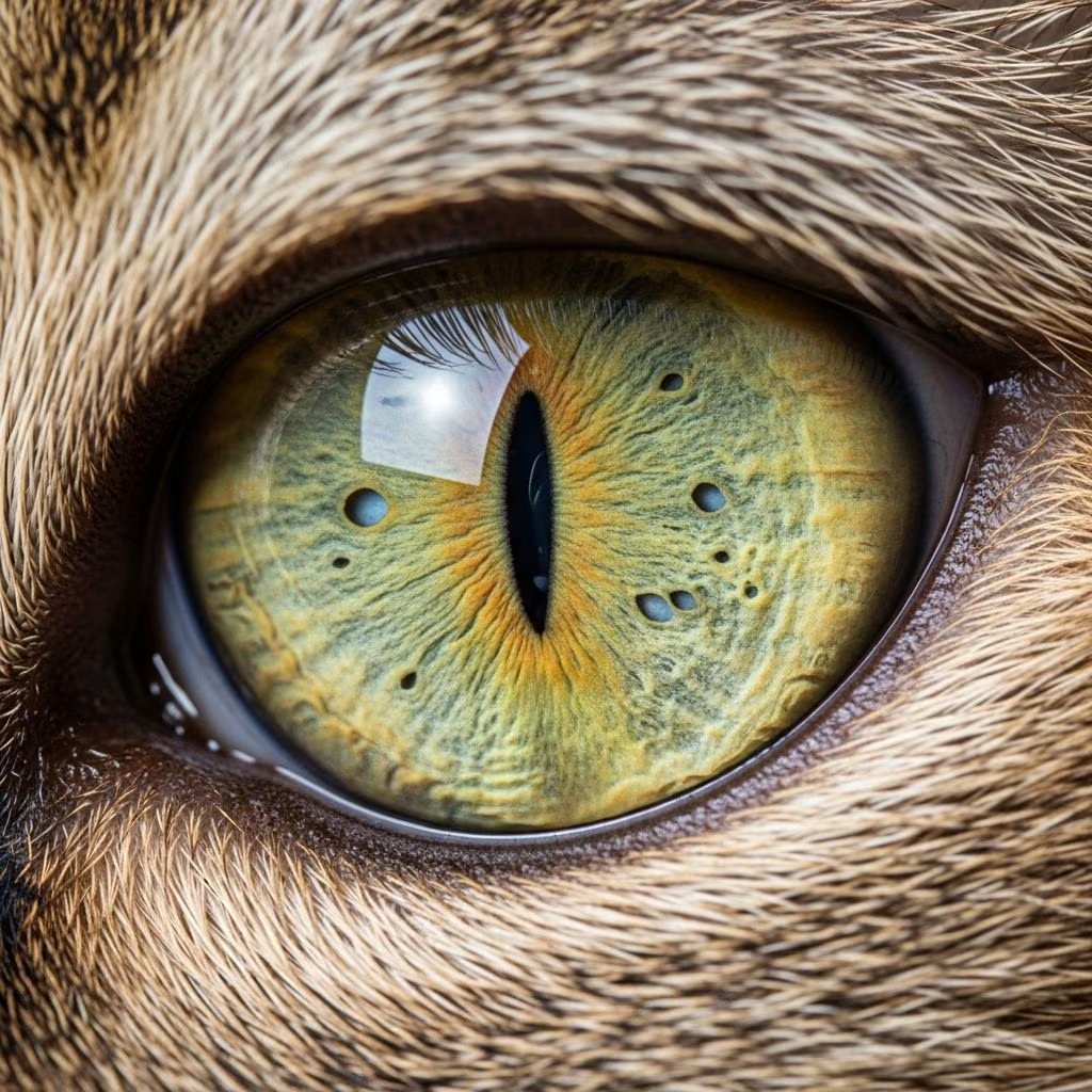
The iris, the colored portion encircling the pupil in a cat’s eye, plays a key role in controlling light entry. Iris atrophy describes the progressive thinning or breakdown of this structure, often noticeable as changes in pupil shape or iris appearance. Primarily observed in senior cats, it can also stem from inflammatory or pressure-related eye disorders. While typically not vision-threatening, it signals potential health concerns requiring veterinary oversight.
Recognizing the Condition: Key Indicators
Early detection hinges on observing subtle eye alterations. Cats with iris atrophy may display several hallmark signs, though these can overlap with other ocular issues.
- Abnormal pupil shapes: Pupils may appear jagged, scalloped, or unevenly sized between eyes, known as anisocoria.
- Light response issues: Pupils fail to constrict fully in bright light, leading to poor adaptation and squinting.
- Visible iris gaps: Thinning creates translucent spots or holes, sometimes mimicking extra pupils.
- Color shifts: Fading or darkening of the iris as underlying layers become exposed.
- Light aversion: Cats seek dim areas or paw at eyes due to discomfort from glare.
These symptoms often develop gradually, prompting owners to mistake them for normal aging. Routine eye checks during wellness visits can catch them early.
Primary Drivers Behind Iris Breakdown
Iris atrophy arises from multiple pathways, with age as the most frequent culprit. Understanding these helps tailor interventions.
Age-Linked Degeneration
In felines over 10 years, senile iris atrophy emerges from natural muscle and tissue wear in the iris sphincter, impairing constriction. This spares vision but heightens photophobia. Blue-eyed breeds show higher incidence, possibly due to pigment scarcity.
Inflammatory Triggers: Uveitis Role
Chronic uveitis, inflammation of the eye’s middle layer, erodes iris tissue over time. Triggers include infections (feline leukemia, toxoplasmosis), immune disorders, or trauma. Persistent cases form scar tissue, exacerbating atrophy.
Pressure Overload from Glaucoma
Elevated intraocular pressure in glaucoma stretches and thins the iris. Untreated, it advances to optic nerve damage and blindness. Secondary atrophy here demands urgent pressure control.
| Cause | Age Group Affected | Key Features | Progression Risk |
|---|---|---|---|
| Senile | Senior (>10 yrs) | Slow, bilateral, non-painful | Low, stable |
| Uveitis-Induced | All ages | Inflammation, pain possible | Moderate, treatable |
| Glaucoma-Related | Variable | High pressure, vision loss | High, urgent |
Diagnostic Steps: Veterinary Evaluation
Vets employ targeted exams to confirm iris atrophy and rule out mimics like congenital defects (iris hypoplasia, coloboma) or polycoria.
- Visual Inspection: Pupil response to light, fluorescein staining for ulcers.
- Tonometry: Measures eye pressure to detect glaucoma.
- Transillumination: Light shone through closed lids reveals iris holes.
- Schirmer Tear Test: Assesses tear production amid inflammation.
- Advanced Imaging: Ultrasound or gonioscopy for structural insights.
Bloodwork or serology may probe systemic causes like infections. Differentiation from neurologic issues ensures accurate management.
Management Strategies: Treatment and Support
Atrophy itself proves irreversible, focusing care on halting progression and easing symptoms. Plans vary by cause.
Targeted Therapies
- Anti-inflammatories: Corticosteroid drops for uveitis to curb swelling.
- Pressure Reducers: Beta-blockers or prostaglandins for glaucoma.
- Antibiotics/Antifungals: If infection drives inflammation.
Avoid mydriatics like atropine, as they worsen sphincter strain.
Home Care Essentials
Complement vet care with daily routines:
- Clean faces gently to avert irritants.
- Apply vet-prescribed lubricants for dry eyes.
- Dim lighting or UV-protective shades indoors.
- Nutrient-rich diets with antioxidants (omega-3s, lutein) support ocular health.
- Monitor for worsening: redness, cloudiness, behavior shifts.
Regular rechecks track stability, adjusting as needed.
Pain and Comfort Considerations
Pure senile atrophy remains painless, but secondary forms from uveitis or glaucoma cause aching, tearing, or rubbing. Prompt intervention prevents escalation. Cats hiding pain subtly necessitate owner vigilance.
Prognosis and Long-Term Outlook
Most cats adapt well, retaining functionality despite cosmetic changes. Senile cases stabilize; treated secondaries improve. Untended glaucoma risks blindness. Lifelong monitoring ensures quality of life.
FAQs on Feline Iris Atrophy
Does iris atrophy blind my cat?
No, it rarely impacts sight directly but flags treatable vision threats like glaucoma.
Can young cats get it?
Rarely; usually secondary to inflammation or injury, not age.
Is surgery an option?
Not typically; meds manage causes effectively.
How to prevent progression?
Annual vet exams, parasite control, prompt injury care.
What breeds are prone?
Blue-eyed cats, though any breed possible.
Enhancing Eye Health Holistically
Beyond specifics, bolster resilience via balanced nutrition, stress reduction, and environmental tweaks. Omega fatty acids from fish oil aid tissue repair; bilberry extracts offer antioxidant protection per studies. Cleanliness curbs bacterial threats. Engage in play to maintain activity, indirectly supporting circulation.
References
- Iris Atrophy in Cats: Signs, Causes & When to See a Vet — Catster. 2023. https://www.catster.com/cat-health-care/iris-atrophy-in-cats/
- Degeneration of the Iris in the Eye in Cats — PetMD. 2023. https://www.petmd.com/cat/conditions/eyes/c_ct_iris_atrophy
- Eye: Iris Atrophy in Cats — Vetlexicon. 2024. https://www.vetlexicon.com/felis/ophthalmology/articles/eye-iris-atrophy/
- EP 450: Iris Atrophy in Cats and Dogs — YouTube (Canine & Feline Medicine). 2025-06-01. https://www.youtube.com/watch?v=_dDKn4k_fdo
- Progressive Retinal Atrophy in Cats — VCA Animal Hospitals. 2024. https://vcahospitals.com/know-your-pet/progressive-retinal-atrophy-in-the-cat



