

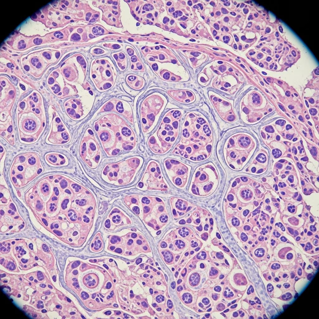
Hyalohyphomycosis represents a group of opportunistic fungal infections caused by hyaline (non-pigmented) molds that primarily affect the skin, subcutaneous tissues, bones, and occasionally disseminate systemically in animals. These infections arise from ubiquitous environmental fungi entering through trauma or inhalation, posing diagnostic and therapeutic challenges in veterinary practice.
Understanding the Fungal Pathogens Behind Hyalohyphomycosis
At the core of hyalohyphomycosis are saprophytic fungi thriving in soil, decaying vegetation, and water sources worldwide. Key genera implicated include Fusarium, Acremonium, Paecilomyces, Pseudallescheria, Scedosporium, and Phialosimplex, which produce transparent hyphae in tissues, distinguishing them from pigmented phaeohyphomycosis agents. These molds are opportunistic, rarely causing disease in healthy hosts but exploiting breaches in skin barriers or immunocompromised states.
In dogs, particularly young large breeds, Fusarium and Paecilomyces species dominate, leading to localized or disseminated forms. Cats experience nodular granulomatous skin lesions most frequently from hyaline fungi, with genera like Sagemonella also noted. Other species, including exotic pets and livestock, face similar risks, though reports are scarcer.
Transmission Routes and Risk Factors
Primary transmission occurs via traumatic implantation, such as puncture wounds from wood splinters, fight bites contaminated with soil, or water exposure. Inhalation of spores can initiate respiratory or central nervous system involvement, though less common. Geographic patterns show higher incidence in tropical and warmer climates, with species variations: Alternaria in Europe and Microsphaeropsis arundinis in Australia.
- Predisposing Factors: Outdoor access, contact with prey or other animals, and environmental exposure heighten risk, even in indoor pets.
- Immunosuppression: Chronic corticosteroid use, FeLV/FIV in cats, or other deficits promote dissemination, though many cases occur in immunocompetent animals.
- Trauma Types: Sharp injuries > bite wounds; rare water contamination.
Clinical Manifestations Across Species
Symptoms vary by site and extent. Localized cutaneous forms present as slow-growing, firm-to-fluctuant nodules, often ulcerated with draining tracts, commonly on the face, nasal planum, pinnae, distal limbs, or tail. In cats, darkly pigmented subcutaneous masses on head and extremities predominate.
| Species | Common Sites | Typical Signs |
|---|---|---|
| Dogs (young, large breeds) | Nose, digits, ears | Nodular lesions, lameness, osteomyelitis |
| Cats | Nares, face, limbs, tail | Ulcerated nodules, neurological deficits |
| Other (e.g., amphibians) | Skin | Discoloration, nodules, shedding |
Disseminated disease manifests with multi-organ involvement: fever, lymphadenopathy, chorioretinitis, or CNS signs like seizures, ataxia, head tilt, and paresis. Acute presentations follow trauma, while chronic cases relapse.
Diagnostic Approaches: From Cytology to Molecular Tools
Early suspicion arises from non-healing wounds or granulomas in at-risk animals. Initial cytology (modified Wright’s stain) reveals pyogranulomatous inflammation with hyphae, though necrotic debris may obscure fungi.
- Histopathology: Gold standard; shows septate hyphae (GMS/PAS stains highlight contours). Biopsy confirms tissue invasion.
- Culture: Essential for speciation; slow-growing molds require fungal media.
- Molecular: PCR with ITS sequencing identifies genera; Aspergillus galactomannan ELISA cross-reacts for screening.
- Imaging: Thoracic/abdominal radiographs for dissemination.
Differentiate from aspergillosis or bacterial abscesses via morphology: hyphae may swell globosely, mimicking Aspergillus.
Treatment Strategies: Surgery Meets Antifungals
Management combines aggressive intervention with prolonged therapy. Localized lesions demand wide-margin surgical excision, often curative if complete.
Systemic Antifungals:
- Itraconazole: First-line; effective for skin/CNS cases, though relapses common.
- Posaconazole/Voriconazole: For resistant/disseminated disease.
- Echinocandins (e.g., Caspofungin): Cell wall inhibitors; promising for hyalohyphomycosis but costly and IV-only.
- Amphotericin B: Reserved for severe cases due to nephrotoxicity.
Immunosuppressed patients require guarded prognoses; monitor via serial cultures/imaging.
Prognosis and Long-Term Outcomes
Excellent for solitary, excisable lesions (>80% cure with surgery + antifungals). Poor for disseminated/multi-lesion cases (high mortality, frequent relapses). Early intervention is key; chronic deep infections resist therapy.
Prevention Measures for Animal Owners and Veterinarians
Minimize trauma in outdoor pets: supervise play, inspect wounds promptly. Avoid corticosteroids without necessity. Routine FeLV/FIV screening in cats aids risk assessment. Environmental control is impractical due to ubiquity, but hygiene post-injury helps.
FAQs on Hyalohyphomycosis in Animals
Q: Is hyalohyphomycosis zoonotic?
A: No documented risk from infected animals to humans.
Q: How long does treatment last?
A: Months; minimum 3-6 months post-resolution, guided by cultures.
Q: Can it affect livestock?
A: Rare reports; similar principles apply, focus on wound care.
Q: What’s the difference from phaeohyphomycosis?
A: Hyalohyphomycosis features clear hyphae; phaeo has pigmented (melanin-rich) forms.
Q: Are there breed predispositions?
A: Young large-breed dogs most affected; no strong cat breeds noted.
Emerging Research and Future Directions
Recent studies emphasize molecular diagnostics for rapid ID and novel azoles/echinocandins for resistance. Veterinary mycology advances promise better outcomes, especially in exotics.
References
- Phaeohyphomycosis and Hyalohyphomycosis in Cats (Felis) — Vetlexicon. 2023. https://www.vetlexicon.com/felis/articles/phaeohyphomycosis-and-hyalohyphomycosis/
- Approaches to Opportunistic Fungal Infections in Small Animals — Today’s Veterinary Practice. 2022-10-01. https://todaysveterinarypractice.com/internal-medicine/approaches-to-opportunistic-fungal-infections-in-small-animals/
- Rare opportunistic mycoses in cats: phaeohyphomycosis and hyalohyphomycosis — PMC/NCBI (Peer-reviewed). 2024. https://pmc.ncbi.nlm.nih.gov/articles/PMC11148967/
- I-F05, Hyalohyphomycosis (paecilomycosis), haired skin, rhesus — AskJPC (Joint Pathology Center). 2023. https://www.askjpc.org/vspo/show_page.php?id=SnMveVBZY3hSZHZuN1M1WGlkZkRnUT09



