

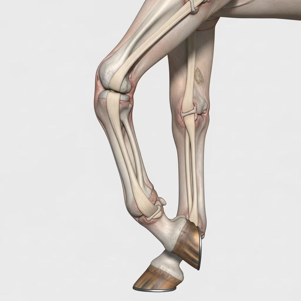
The forelimb of a horse plays a critical role in locomotion, with the carpus—commonly called the knee—and the metacarpus, or cannon bone region, bearing significant weight and stress during movement. Disorders in these areas can lead to lameness, reduced performance, and long-term complications if not addressed promptly. Understanding the anatomy, recognizing early signs, and knowing treatment options are vital for horse owners, trainers, and veterinarians.
Anatomy of the Equine Forelimb: Focus on Carpus and Metacarpus
The equine carpus consists of three primary joints: the antebrachiocarpal, middle carpal, and carpometacarpal joints. These are supported by two rows of cuboidal carpal bones positioned between the radius and the metacarpal bones. Intercarpal ligaments and dorsal ligaments connect these bones, enabling primarily flexion and extension motions.
The metacarpus includes the prominent third metacarpal bone, known as the cannon bone, flanked by the smaller second and fourth metacarpal splint bones. Evolutionarily, the horse’s forelimb has reduced to one main weight-bearing digit (III), with splint bones representing remnants of digits II and IV. This structure provides stability but is prone to specific injuries due to high-speed impacts and repetitive strain.
- Antebrachiocarpal joint: Allows major flexion and extension.
- Middle carpal joint: Communicates with the carpometacarpal joint, contributing to smooth motion.
- Carpometacarpal joint: Offers limited movement, linking carpal bones to metacarpals.
Ligaments, tendons like the extensor carpi radialis, and the carpal canal with its enclosed structures further stabilize the region, preventing excessive flexion during standing or gait.
Common Signs of Carpus and Metacarpus Disorders
Horses with issues in these areas often exhibit noticeable lameness, particularly evident at trot. Swelling, joint effusion (fluid buildup), heat, and pain on palpation are hallmark signs. Lameness may worsen with flexion tests or circling, and in metacarpal cases, deformities or crepitus (grating sounds) may occur.
| Symptom | Carpus Indicators | Metacarpus Indicators |
|---|---|---|
| Lameness Grade | Mild to severe, often bilateral | Acute, sharp on weight-bearing |
| Swelling | Joint distension, soft tissue | Localized to bone or splint |
| Pain Response | Positive to flexion/extension | Tender on direct pressure |
| Other | Instability, buckling | Deformity, exostosis |
Early detection through consistent lameness exams can prevent progression to chronic conditions.
Key Disorders Affecting the Carpus
Chip Fractures and Osteochondrosis
Chip fractures, often from trauma in racing or jumping horses, involve small bone fragments in the carpal joints. Osteochondrosis dissecans (OCD) arises from abnormal cartilage development, leading to flaps or loose bodies that cause inflammation and lameness. Young horses aged 1-2 years are particularly susceptible.
Symptoms include intermittent lameness and joint effusion. Radiographs reveal fragments, while arthroscopy confirms diagnosis and allows fragment removal.
Synovial Herniations and Tears
Full-thickness tears in the joint capsule or synovial pad can lead to herniations, where synovium protrudes, causing pain and locking sensations. These are common in performance horses due to hyperextension injuries.
Degenerative Joint Disease (DJD)
Chronic wear leads to osteoarthritis, with cartilage erosion, bone remodeling, and periarticular osteophytes. Progression results in persistent lameness and reduced range of motion.
Metacarpus-Specific Conditions
Splint Bone Issues
Exostoses (bony proliferations) on splint bones arise from ligament strain or direct trauma. The second splint is more prone due to its position. Inflammation causes heat and swelling, potentially impinging on the cannon bone.
Cannon Bone Fractures
Complete or incomplete fractures of the third metacarpal range from stress fractures in endurance horses to catastrophic breaks in racers. Condylar fractures propagate proximally, demanding surgical intervention like lag screw fixation.
Buck Shins and Saucer Fractures
Dorsal metacarpal disease, or buck shins, involves periosteal reaction in young Thoroughbreds from high-speed training. Saucer fractures are shallow dorsal cortex defects.
Management includes rest, shockwave therapy, and monitoring via scintigraphy for early stress reactions.
Diagnostic Approaches
Diagnosis starts with clinical exam, including hoof testers, flexion tests, and gait analysis. Imaging is cornerstone:
- Radiography: Standard views (lateral, dorsopalmar, obliques) detect fractures, OCD, and DJD.
- Ultrasonography: Assesses soft tissues, ligaments, and tendon sheaths.
- Scintigraphy: Identifies stress fractures via bone uptake.
- MRI/CT: Advanced for subtle cartilage or subchondral issues.
- Arthroscopy: Diagnostic and therapeutic for intra-articular pathology.
Flexion tests specifically load the carpus, exacerbating lameness if affected.
Treatment Strategies
Treatments vary by condition severity:
- Conservative: Stall rest (4-6 weeks), NSAIDs (phenylbutazone), cold therapy, and controlled exercise for minor strains or buck shins.
- Intra-articular: Corticosteroids or hyaluronic acid for synovitis/OCD.
- Surgical: Arthroscopic debridement for chips/OCD; fracture repair with screws/plates; splint bone excision for exostoses.
- Regenerative: Stem cells, PRP (platelet-rich plasma), or IRAP for joint repair.
Post-treatment rehab involves gradual return to work, monitored by serial imaging.
Prevention and Management Tips
Proactive measures reduce incidence:
- Balanced nutrition with proper calcium:phosphorus ratios to support bone growth.
- Gradual conditioning programs, avoiding overtraining in juveniles.
- Proper footing and shoeing to minimize torque.
- Routine lameness screening in performance horses.
- Vaccinations and deworming to prevent systemic contributors to joint health.
Farriers play a key role in corrective shoeing for angular limb deformities affecting carpus loading.
Prognosis and Long-Term Outlook
Prognosis depends on lesion type and timeliness of intervention. Simple chip fractures carry good outcomes (80-90% return to work), while severe comminuted fractures or advanced DJD may lead to retirement. Regular monitoring ensures early detection of recurrence.
Frequently Asked Questions (FAQs)
What causes sudden lameness in a horse’s knee?
Sudden carpal lameness often stems from chip fractures, ligament tears, or synovitis from trauma. Immediate vet evaluation is crucial.
Can cannon bone fractures heal without surgery?
Non-displaced stress fractures may heal with rest and therapy, but complete fractures typically require surgical stabilization.
How long does recovery take for carpal OCD?
Post-arthroscopy recovery spans 3-6 months, with gradual exercise resumption.
Are splint bone lumps always problematic?
Not always; many are cosmetic, but painful or progressive ones need attention to prevent interference.
What’s the best way to prevent buck shins?
Controlled training on soft surfaces, proper warm-up, and bone scans in at-risk horses.
References
- The Equine Carpus — Veterian Key. 2023. https://veteriankey.com/the-equine-carpus/
- Equine Carpus Anatomy — SlideShare. 2015-01-15. https://www.slideshare.net/slideshow/carpal-anatomy/42896775
- Thoracic Limb – Forelimb — University of Minnesota Pressbooks. 2023. https://pressbooks.umn.edu/largeanimalanatomy/chapter/thoracic-limb-forelimb/
- The evolution and anatomy of the horse manus: Digit reduction — PMC (NCBI). 2018-02-14. https://pmc.ncbi.nlm.nih.gov/articles/PMC5792948/
- Anatomy of the Horse’s Metacarpal Bone and Digit — YouTube (Veterinary Channel). 2023. https://www.youtube.com/watch?v=hUfwUvCfX5w
- Disorders of the Carpus and Metacarpus in Horses — Merck Veterinary Manual. 2023. https://www.merckvetmanual.com/horse-owners/bone-joint-and-muscle-disorders-in-horses/disorders-of-the-carpus-and-metacarpus-in-horses
- Equine Forelimb Carpus Example 1 — University of Illinois VetMed. 2023. https://vetmed.illinois.edu/imaging_anatomy/equine/forelimb/carpus/ex01/carpus01-eq.html



