

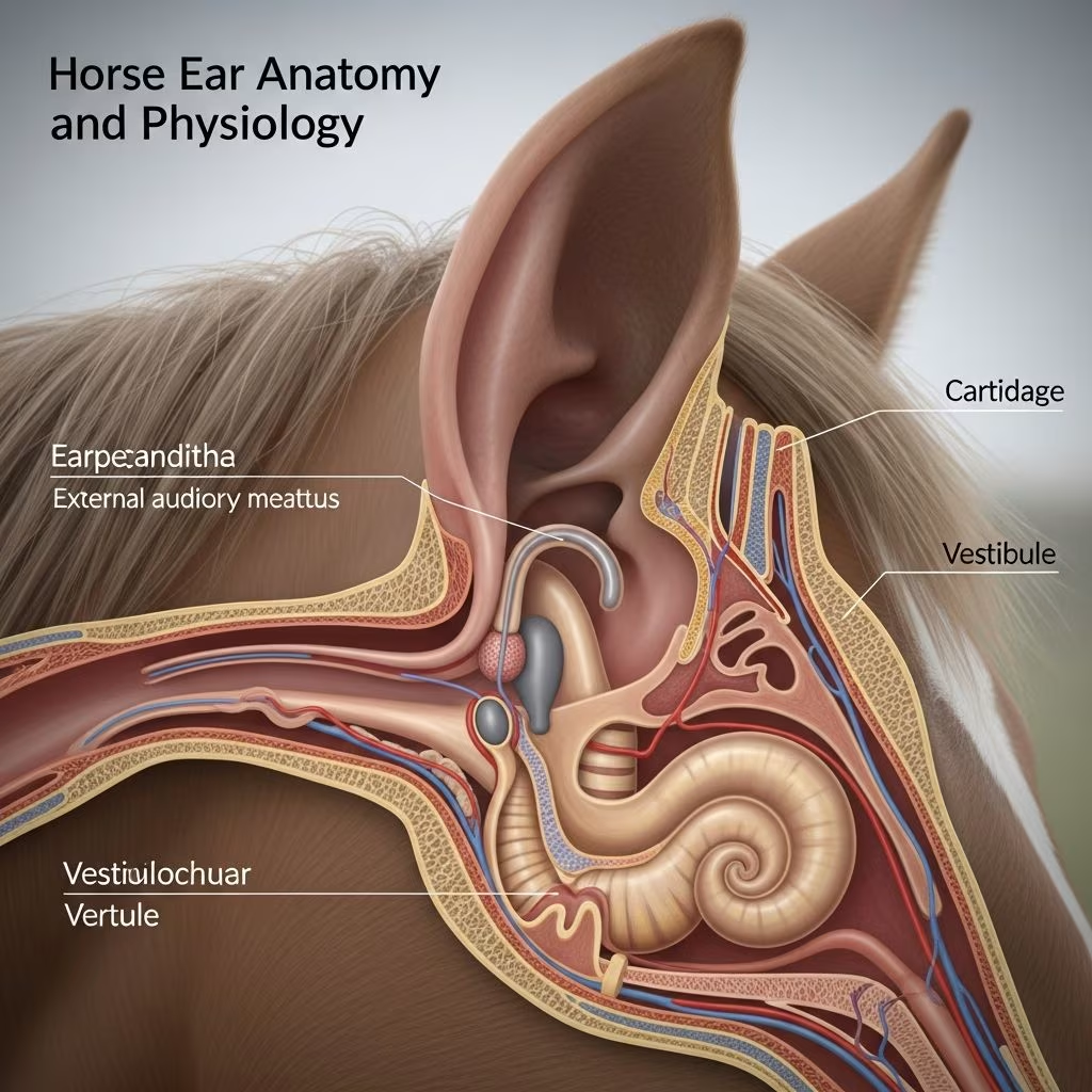
The equine ear is a sophisticated sensory organ essential for a horse’s survival, enabling precise hearing, equilibrium maintenance, and nonverbal signaling. Comprising outer, middle, and inner sections, it processes auditory signals and spatial orientation with remarkable efficiency, adapted to the horse’s prey animal instincts.
The Outer Ear: Gateway to Sound Capture
The visible portion of the horse’s ear, known as the pinna or auricle, serves as a dynamic collector of sound waves. This funnel-like structure, primarily composed of flexible cartilage covered by thin skin, amplifies faint noises crucial for detecting predators. Its large size and mobility allow horses to pinpoint sound sources across a wide field without head movement.
Supported by 10 pairs of muscles, the pinna rotates up to 180 degrees independently, facilitating nearly 360-degree auditory coverage. This muscular control not only enhances hearing but also acts as a visual cue: forward-pointing ears indicate curiosity, while flattened ones signal aggression or fear.
Extending inward, the external ear canal is a long, narrow tube lined with haired skin and abundant sebaceous glands. In horses, this canal transitions from cartilaginous to bony portions, with an abrupt shift in epithelial pigmentation marking the junction. The elongated design protects deeper structures from debris while funneling vibrations to the tympanic membrane.
- Key features of the outer ear:
- Cartilage-dominated pinna for flexibility and shape retention.
- Muscular attachments enabling precise positioning.
- Long canal preventing easy visualization of the eardrum, unlike in smaller animals.
Middle Ear Mechanics: Amplifying Vibrations
At the canal’s terminus lies the tympanic membrane, a delicate, obliquely oriented drum that vibrates upon sound impact. This membrane anchors the middle ear’s ossicles: malleus (hammer), incus (anvil), and stapes (stirrup), the body’s smallest bones linked in a chain.
The malleus attaches directly to the membrane, transmitting motion to the incus and then the stapes, which contacts the oval window—a gateway to the inner ear. Horses exhibit species-specific ossicle variations, including a prominent tensor tympani muscle that tenses the membrane to dampen loud noises and protect inner structures.
The middle ear cavity, or tympanic bulla, connects to the nasopharynx via the auditory tube, equalizing pressure and draining secretions. Muscles like the tensor veli palatini, innervated by cranial nerves, regulate this tube’s patency. Histological studies confirm these components mirror mammalian norms but feature unique traits, such as robust annular cartilage bridging to the bony canal.
| Ossicle | Function | Horse-Specific Note |
|---|---|---|
| Malleus | Attaches to tympanic membrane | Long handle for efficient vibration transfer |
| Incus | Links malleus to stapes | Short and long crura for stability |
| Stapes | Presses on oval window | Compact footplate suited to equine canal |
Inner Ear: Hearing and Balance Nexus
The inner ear houses the cochlea for audition and vestibular apparatus for balance, forming a fluid-filled bony labyrinth. Sound-induced stapes motion creates fluid waves in the scala media, stimulating hair cells along the basilar membrane to generate nerve impulses via the cochlear nerve.
Adjacent semicircular canals detect angular acceleration, while utricle and saccule sense linear motion, feeding the vestibular nerve for proprioception. This dual system ensures horses maintain agility during flight responses, with thousands of sensory cells providing high-fidelity signals.
Histology reveals equine inner ear congruence with humans, including specialized rete pegs in the canal and a thick tensor tympani, underscoring adaptations for open environments. Damage here can cause not only deafness but also ataxia, circling, or head tilt.
Sensory Roles Beyond Basic Hearing
Horse ears excel in low-frequency detection, vital for long-distance herd calls and environmental monitoring. Their 360-degree capability, aided by pinna swivel, surpasses human directional hearing, enhancing predator evasion.
Equilibrium functions prevent disorientation in uneven terrain, while ear positioning communicates intent: pricked forward for focus, swiveled back for rear alerts. These behaviors reflect neural integration of auditory and vestibular inputs with cerebral processing.
Clinical Relevance and Health Maintenance
Understanding ear anatomy aids in spotting issues like otitis externa (canal inflammation from insects/debris), media (ossicle infections), or interna (labyrinthitis from temporohyoid joint degeneration). Symptoms include head shaking, discharge, or balance loss; early vet exams are crucial given the canal’s inaccessibility.
Preventive care involves fly masks, clean environments, and routine checks for ticks/parasites. Nutritional support for cartilage health (e.g., omega-3s) bolsters resilience. Advanced imaging like CT scans reveal osseous changes in chronic cases.
- Common ear threats:
- Insect bites causing secondary infections.
- Trauma from halters or fights.
- Guttural pouch mycosis eroding nearby structures.
Behavioral Insights from Ear Posture
Ears are equine mood barometers: neutral flop signals relaxation, rigid forward attention, pinned aggression. Independent movement amplifies selective listening, conserving energy. Trainers leverage this for assessing stress during riding or handling.
In herds, synchronized ear flicks coordinate alerts, reinforcing social bonds. Deviations, like persistent droop, may indicate pain or neurology, warranting intervention.
Comparative Anatomy Highlights
Versus dogs/cats, horse canals are tubular and deep, complicating otoscopy. Ossicles are scaled for body size, with stronger damping muscles. Inner ear density supports high-speed maneuvers, differing from slower grazers.
FAQs on Horse Ear Anatomy
Why are horse ears so mobile?
Horse ears have 10 muscles per pinna, allowing 180-degree rotation for panoramic hearing and communication.
Can you see a horse’s eardrum easily?
No, the long, angled canal obscures it, unlike in small animals.
What causes head tilting in horses?
Often vestibular issues in the inner ear or temporohyoid joint problems affecting balance.
How do horses hear better than humans?
Larger pinnae amplify sound, and they detect wider frequencies, especially lows.
Is ear cartilage prone to injury?
Yes, flexible but vulnerable to tears from fights or ill-fitting tack; heals slowly due to poor vascularity.
Advanced Research Directions
Ongoing studies use histology to map sensory cell distributions, aiding therapies for age-related loss. Genetic analyses explore breed variations, like draft horses’ upright ears versus mules’ droops. Biomechanics models simulate vibration transmission, informing prosthetic designs.
References
- The Anatomy of the Ear — The Horse. 2014-approx. https://thehorse.com/14457/the-anatomy-of-the-ear/
- The Ear and Eye — Veterian Key. 2016-approx. https://veteriankey.com/the-ear-and-eye/
- Histological study of the external, middle and inner ear of horses — PubMed (Anat Histol Embryol). 2014-10-07. https://pubmed.ncbi.nlm.nih.gov/25283481/
- Listen Up: The Fascinating World of Horse Ears — EquiMade. 2023-approx. https://www.equimade.com/listen-up-the-fascinating-world-of-horse-ears/
- Parts of a Horse: Horse Anatomy with Pictures — Farmhouse Tack. 2022-approx. https://www.farmhousetack.com/blogs/barn-blog/parts-of-a-horse-horse-anatomy-with-pictures-equestrians-guide



