

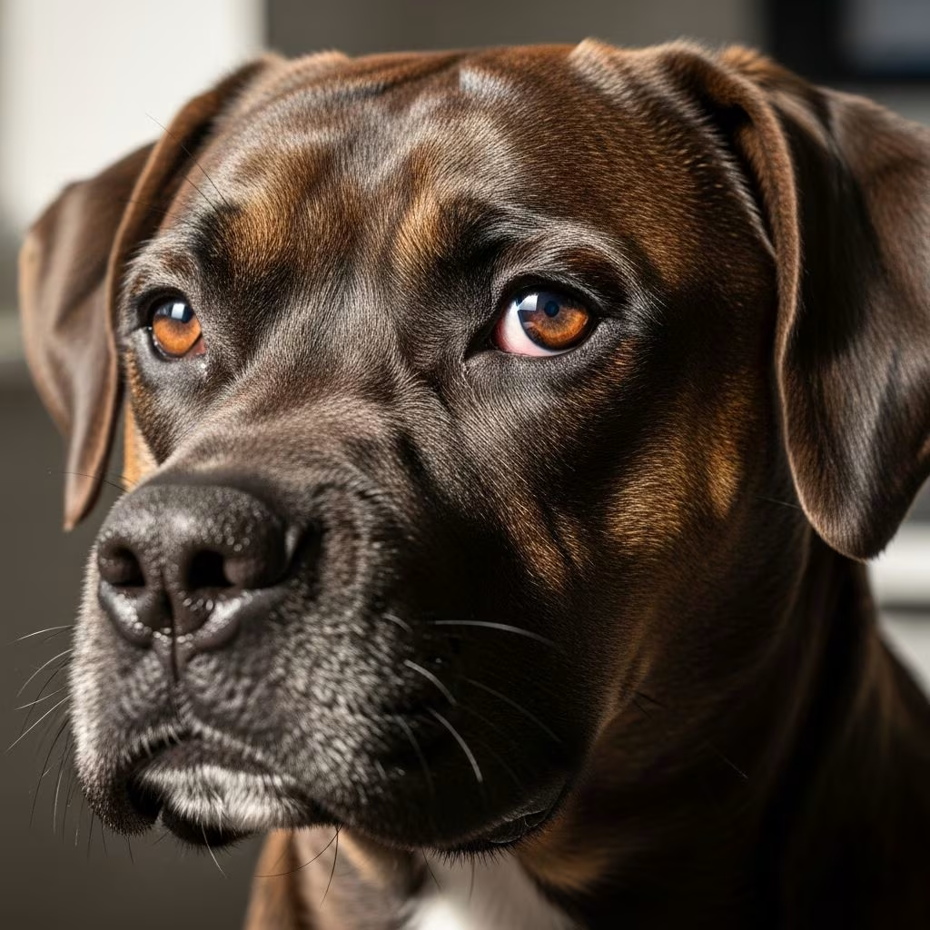
Understanding Horner’s Syndrome in Dogs
Horner’s syndrome is a common neurological disorder that affects the eye and facial muscles in dogs. This condition typically occurs suddenly and is caused by interruption of the sympathetic nerve pathway that supplies the eyes and facial structures. Understanding this condition is crucial for dog owners, as early recognition and proper veterinary intervention can significantly improve outcomes for affected pets.
The syndrome results from damage to the sympathetic nervous system, which normally works in balance with the parasympathetic nervous system to maintain proper eye function and facial muscle tone. When the sympathetic innervation to the eye is interrupted, dogs develop a characteristic collection of clinical signs that veterinarians can readily identify.
Clinical Signs and Symptoms
Horner’s syndrome presents with a distinctive set of ocular and facial symptoms that develop suddenly in affected dogs. These signs result from the loss of sympathetic nerve function to the eye and surrounding structures. The most commonly observed clinical manifestations include:
- Miosis (Constricted Pupil): The pupil on the affected side becomes noticeably smaller than the pupil on the unaffected side, creating an obvious asymmetry when comparing both eyes.
- Ptosis (Drooping Eyelid): The upper eyelid droops due to loss of muscle tone, giving the affected eye a sleepy or partially closed appearance.
- Enophthalmos (Sunken Eye): The eyeball appears to retract back into the socket, making the eye look shrunken or recessed.
- Protrusion of the Third Eyelid: The nictitating membrane (third eyelid) becomes elevated and more visible due to the sunken appearance of the eyeball.
- Increased Warmth and Pink Coloration: The nose and ear on the affected side may appear warmer and display increased pink coloration compared to the unaffected side.
In some cases, dogs may also experience additional symptoms such as excessive salivation or difficulty eating on the affected side. It’s important to note that Horner’s syndrome typically affects only one eye and facial side. If bilateral involvement occurs, it suggests underlying brainstem disease, which requires immediate veterinary attention.
Causes of Horner’s Syndrome
Horner’s syndrome can develop from various underlying conditions that damage the sympathetic nerve pathway. The sympathetic nerves responsible for normal eye and facial muscle function extend from the brain through the spinal cord and into the peripheral nervous system.
Identified Causes
When a specific cause can be determined, common underlying conditions include:
- Trauma and Injury: Head, neck, or thoracic trauma from accidents, falls, or bites can damage the sympathetic nerves.
- Ear Infections: Middle or inner ear infections (otitis media/interna) are among the most common identified causes of Horner’s syndrome.
- Tumors and Neoplasms: Brain tumors, brainstem tumors, or thoracic tumors can compress or damage sympathetic nerve pathways.
- Surgical Procedures: Ear canal ablation, bulla osteotomy, thoracic surgery, or anesthesia-related complications can lead to nerve damage.
- Intervertebral Disc Disease: Spinal cord compression from disc herniation can interrupt nerve pathways.
- Infectious Diseases: Certain infections such as tick paralysis or neosporosis have been associated with Horner’s syndrome.
- Diabetes Mellitus: Though rare, diabetes has been reported as an underlying cause.
- Vascular Events: Fibrocartilaginous embolism and other vascular events can damage neural tissue.
Idiopathic Cases
Remarkably, approximately 50% of Horner’s syndrome cases in dogs are classified as idiopathic, meaning no specific underlying cause can be identified despite thorough diagnostic investigation. In these cases, the syndrome often resolves spontaneously within 16 weeks to 6 months, suggesting a self-limiting nature of the condition. Golden Retrievers appear to have a higher predisposition to idiopathic Horner’s syndrome compared to other breeds.
Diagnostic Procedures and Tests
Accurate diagnosis of Horner’s syndrome involves a combination of clinical examination and specialized diagnostic testing. The goal is not only to confirm the diagnosis but also to identify any underlying cause requiring treatment.
Initial Examination
Veterinarians begin with a thorough eye and ear examination, looking for signs consistent with Horner’s syndrome and evaluating for obvious causes such as ear infections or masses. The characteristic constellation of clinical signs—constricted pupil, drooping eyelid, sunken eye, and elevated third eyelid—typically confirms the diagnosis.
Pharmacologic Tests
Specialized eye drops are used to help localize the lesion and confirm Horner’s syndrome:
- Cocaine Test: Application of a 5–10% cocaine solution is considered the gold standard for diagnosing Horner’s syndrome. In healthy dogs, cocaine dilates the pupil, whereas affected dogs show little to no pupil dilation. This test is particularly useful in confirming the diagnosis.
- Apraclonidine Test: This 0.5–1% solution has minimal effect on pupil size in healthy eyes but causes pupil dilation in affected eyes, reducing the asymmetry between pupils within 30–45 minutes.
- Phenylephrine or Topical Adrenaline: These medications help determine the specific location of the lesion along the sympathetic nerve pathway, allowing for more targeted diagnostic investigation.
Advanced Imaging
When an obvious cause isn’t apparent, advanced diagnostic imaging may be recommended:
- X-rays (Radiographs): Skull and chest radiographs help identify tumors, trauma, or other structural abnormalities.
- CT Scans: Computed tomography provides detailed cross-sectional images useful for detecting lesions in the brain, brainstem, or spinal cord.
- MRI: Magnetic resonance imaging offers superior soft tissue detail and is particularly useful for identifying nerve damage, tumors, and other neurological abnormalities.
Treatment Options
Treatment of Horner’s syndrome depends on whether an underlying cause has been identified. Since Horner’s syndrome itself is not painful and does not significantly affect vision, the primary treatment goal focuses on addressing any underlying disease process.
Managing Underlying Causes
When a specific cause is identified, targeted treatment becomes the priority:
- Infections: Bacterial ear infections are treated with antimicrobial medications, including oral antibiotics and topical ear treatments as appropriate.
- Tumors: Treatment may involve surgery, chemotherapy, or other specialized cancer therapies depending on the tumor type and location.
- Neurological Conditions: Intervertebral disc disease may require surgical intervention, while other conditions may benefit from anti-inflammatory medications or supportive care.
- Vestibular Disease: Anti-nausea medications such as Cerenia may be prescribed to address accompanying symptoms.
Symptomatic Treatment
Phenylephrine eye drops can be applied topically to stimulate sympathetic nervous system function and help restore normal pupil size and eyelid position. However, these drops are reserved for cases where the elevated third eyelid interferes with vision, as the drops can potentially irritate the cornea. Treatment with phenylephrine is generally considered more of a cosmetic resolution than a curative treatment.
Alternative Therapies
Recent case reports suggest that acupuncture may have a role in managing idiopathic Horner’s syndrome. Some dogs have shown remarkable improvement following acupuncture treatment, with complete resolution of symptoms occurring within days. While research is limited, acupuncture represents a non-invasive option worth discussing with your veterinarian.
Prognosis and Natural Recovery
The prognosis for dogs with Horner’s syndrome varies based on the underlying cause. Most cases of Horner’s syndrome resolve spontaneously without specific treatment. Dogs with idiopathic Horner’s syndrome typically recover completely within 16 weeks to 6 months, depending on the severity of clinical signs.
For dogs with an identified underlying cause, the prognosis depends on the nature and treatability of that condition. Some conditions, such as ear infections, respond well to appropriate medical treatment and may result in complete resolution of Horner’s syndrome. Other conditions, such as certain types of tumors or brainstem disease, may carry a more guarded prognosis.
Regular follow-up veterinary examinations are essential to monitor progress, adjust treatment as needed, and ensure your dog’s overall health and comfort throughout the recovery process.
Ongoing Care and Support
Supporting your dog through Horner’s syndrome involves maintaining comfort and adhering closely to your veterinarian’s treatment recommendations. Keep appointments for scheduled check-ups to allow your vet to monitor progress and make any necessary adjustments to the treatment plan.
While Horner’s syndrome itself is not painful or vision-threatening, the underlying cause may require ongoing management. Continue prescribed medications exactly as directed, and report any changes in your dog’s condition to your veterinarian promptly.
Frequently Asked Questions
Q: Is Horner’s syndrome painful for dogs?
A: No, Horner’s syndrome itself is not painful and does not affect your dog’s vision. However, the underlying cause may require treatment and management.
Q: Can Horner’s syndrome affect both eyes?
A: While extremely rare, bilateral Horner’s syndrome can occur and typically indicates underlying brainstem disease, which requires immediate veterinary evaluation.
Q: How long does it take for Horner’s syndrome to resolve?
A: Most cases resolve spontaneously within 16 weeks to 6 months. However, some cases may resolve more quickly or persist longer depending on the underlying cause.
Q: Will my dog’s appearance return to normal?
A: In most cases, yes. As the sympathetic nerve function recovers, the clinical signs gradually resolve and your dog’s appearance returns to normal.
Q: What should I do if I notice signs of Horner’s syndrome?
A: Contact your veterinarian promptly for evaluation. Early diagnosis and treatment of any underlying cause can improve outcomes and ensure your dog’s health and comfort.
References
- Horner’s Syndrome in Dogs — VCA Animal Hospitals. 2024. https://vcahospitals.com/know-your-pet/horners-syndrome-in-dogs
- Treatment for Horner’s Syndrome in Dogs — PetMD. 2024. https://www.petmd.com/dog/conditions/neurological/horners-syndrome-dogs
- Horner’s Syndrome in Dogs: Causes, Symptoms, Treatment — Integrative Veterinary Care. 2024. https://toegrips.com/horners-syndrome-dogs/
- Horner’s syndrome signs in dogs — Veterinary Vision Center. 2024. https://veterinaryvisioncenter.com/horners-syndrome-in-dogs/
- Horner’s syndrome in dogs: aetiology and diagnostic protocol — Vets and Clinics. 2024. https://vetsandclinics.com/en/horners-syndrome-in-dogs-aetiology-and-diagnostic-protocol
- Horner’s Syndrome in Dogs – Veterinary Teaching Hospital — Colorado State University College of Veterinary Medicine and Biomedical Sciences. 2024. https://vetmedbiosci.colostate.edu/vth/services/ophthalmology/horners-syndrome/



