

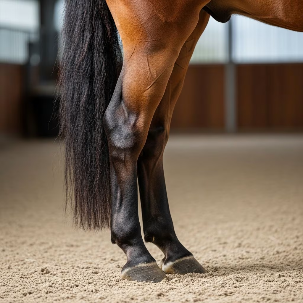
The tarsal region, commonly referred to as the hock, represents one of the most heavily used joints in a horse’s hindlimb and consequently one of the most prone to injury and degenerative conditions. This complex anatomical structure experiences significant biomechanical stress during locomotion, making it susceptible to various pathological conditions that can compromise performance and soundness. Understanding these disorders is essential for horse owners and caretakers seeking to maintain their animals’ health and athletic capability.
Anatomical Foundation of the Tarsal Region
The hock comprises multiple bones and articulations working in coordinated fashion to enable hindlimb movement and shock absorption. The tarsal region contains four distinct synovial compartments, each with specific functions in weight-bearing and locomotion. These compartments include the tarsocrural joint, which connects the tibia to the primary tarsal bones, and the more distal joints that involve the smaller tarsal bones and metatarsal structures.
The biomechanical demands placed on the tarsus during various gaits explain why pathological changes frequently develop in this region. During stance phase, compression forces concentrate on specific tarsal structures, while swing phase requires extensive flexion and extension movements. The presence of specialized locking mechanisms among the talus, central tarsal bone, and third tarsal bone provides stability during weight-bearing but also predisposes certain articulations to excessive wear patterns.
Primary Categories of Tarsal Joint Disease
Degenerative Joint Changes and Arthritis
Osteoarthritis of the tarsal region represents the most commonly encountered disorder affecting the hock. This degenerative condition develops progressively as articular cartilage deteriorates and bone remodeling occurs in response to chronic inflammation and mechanical stress. Horses experiencing tarsal arthritis typically demonstrate reduced hindlimb engagement, diminished push-off capability, and visible changes in movement quality.
The development of arthritis often correlates with prior joint trauma, biomechanical conformational issues, or repetitive high-impact activities. Radiopharmaceutical studies in sound horses reveal highest uptake in dorsal and lateral distal tarsal regions, indicating these areas experience significant adaptive bone modeling in response to loading forces. When protective mechanisms become overwhelmed, cartilage damage and inflammatory changes accelerate.
Bone Spavin and Intertarsal Fusion
Bone spavin represents a specific form of tarsal arthritis characterized by bone proliferation and eventual fusion of distal tarsal joints. This condition develops as the body attempts to stabilize damaged joint surfaces through new bone formation. While bone spavin may eventually reach a chronic phase where lameness stabilizes or improves, the initial formation period typically causes significant discomfort and performance limitations.
The progression of bone spavin varies considerably among horses. Some animals may compensate effectively and return to work after the initial acute phase, while others develop increasingly severe lameness. Advanced imaging techniques help determine the extent of joint involvement and predict long-term functional outcomes.
Bog Spavin and Synovial Distension
Bog spavin refers to fluid distension of the tarsal joint capsule, visible as soft swelling on the medial and dorsomedial aspects of the hock. Longitudinal swelling from joint capsule distension represents a common incidental finding in both sound and lame horses, occurring unilaterally or bilaterally. This condition alone rarely associates with clinical lameness but may indicate underlying joint inflammation or damage.
The presence of bog spavin without concurrent lameness often reflects the body’s response to joint stress rather than active pathological disease. However, progressive distension or associated pain responses warrant diagnostic investigation to exclude other hock pathologies.
Clinical Presentation and Diagnostic Identification
Gait Alterations and Performance Changes
Horses with tarsal disorders frequently display subtle changes in movement quality before overt lameness becomes apparent. Affected animals may demonstrate reduced tarsal flexion during stance phase and decreased forward sliding of distal joints, resulting in diminished propulsion and altered gait mechanics. The appearance shifts toward less bounce and elasticity in the stride, particularly when the horse performs movements requiring maximum tarsal flexion.
Performance-related changes often precede detectable lameness by weeks or months. Owners may notice reduced engagement of the hindquarters, difficulty maintaining speed, or reluctance to perform specific movements such as tight turns, backing, or jumping. Subtle gait deficiencies frequently go unrecognized until lameness becomes more obvious.
Locating Pain Through Palpation
Physical examination of the hock involves systematic palpation of tarsal structures and specific response tests. Compression of the distal medial tarsus and proximal medial metatarsal region often elicits positive responses in horses with tarsal dysfunction, with dynamic testing involving compression followed by trotting exacerbating lameness. This targeted compression identifies pain localization before subjecting the horse to exercise.
However, pain localization through diagnostic analgesia frequently reveals involvement of the distal hindlimb rather than superficial anatomical landmarks, emphasizing the complex pain referral patterns associated with tarsal pathology. Multiple injection sites may be necessary to definitively identify the primary source of discomfort.
Imaging Investigation Protocols
Radiographic evaluation remains the primary diagnostic tool for assessing tarsal pathology. Multiple views including lateral, dorsopalmar, and oblique projections enable visualization of bone proliferation, joint space narrowing, and other degenerative changes. Some conditions may only appear visible in specific radiographic planes, necessitating comprehensive imaging protocols.
Advanced imaging modalities including nuclear scintigraphy and ultrasound provide additional diagnostic information. Scintigraphy reveals areas of increased metabolic activity, helping identify sites of bone remodeling or inflammation not visible on radiographs. Ultrasound examination proves particularly valuable for evaluating soft tissue structures including ligaments, tendons, and joint capsules.
Understanding Specific Tarsal Conditions
Osteochondrosis of the Distal Tarsus
Osteochondrosis represents a developmental orthopedic disease affecting the growth cartilage of tarsal bones. Osteochondral lesions occurring in small tarsal bones may predispose to subsequent distal tarsal osteoarthritis, though the precise relationship between initial vascular failure and long-term degenerative changes remains incompletely understood. These lesions range from subclinical findings to significant sources of lameness depending on lesion characteristics and location.
Young horses with incomplete ossification of cuboidal tarsal bones face particular risk for chronic lameness and reduced athletic ability. The timing of bone maturation and degree of complete ossification correlate with performance capacity, making early detection of developmental abnormalities increasingly important for athletic horses.
Collateral Ligament Dysfunction
The tarsal collateral ligaments provide critical stabilization during weight-bearing and movement. Short medial collateral ligaments bear responsibility for the snap-joint phenomenon observed during tarsocrural motion, providing dynamic stability through their fiber arrangement and anatomical positioning. Injury to these structures compromises joint stability and predisposes to secondary arthritic changes.
Collateral ligament injuries may result from direct trauma, sudden torque forces, or chronic overstress from poor conformation or inappropriate training. Diagnosis requires careful evaluation of collateral ligament integrity through palpation and diagnostic imaging.
Superficial Digital Flexor Tendon Complications
The superficial digital flexor tendon passes over the calcaneal tuberosity, making it vulnerable to strain and luxation in the tarsal region. While tendon lesions can be identified ultrasonographically, they frequently remain unassociated with lameness. However, significant tendon damage or displacement may contribute to joint dysfunction and pain.
Management Approaches and Treatment Considerations
Conservative Management Strategies
Initial management of many tarsal disorders focuses on anti-inflammatory therapy and activity modification. Nonsteroidal anti-inflammatory medications reduce pain and inflammation, enabling continued controlled movement that maintains joint mobility. Restricted exercise protocols balance the need for activity with protection from excessive stress.
Intra-articular corticosteroid injections provide localized anti-inflammatory effects within the tarsal joint capsule. These treatments offer variable efficacy depending on underlying pathology severity and joint compartment involvement. Repeated injections must be carefully considered against potential long-term effects on cartilage and bone.
Diagnostic Analgesia and Treatment Response
Diagnostic joint injections serve dual purposes by identifying the primary site of discomfort while simultaneously delivering therapeutic medication directly to affected structures. Response to diagnostic analgesia guides treatment decisions and helps predict long-term prognosis. Horses demonstrating improved lameness following anesthesia of specific compartments likely benefit from targeted intervention at those sites.
The variable nature of tarsal pain patterns necessitates comprehensive diagnostic protocols before commencing treatment. Multiple joint compartments may require evaluation, as disease in proximal compartments can affect distal structures and create complex pain patterns.
Regenerative and Advanced Therapeutic Options
Emerging therapeutic approaches including platelet-rich plasma, stem cell therapies, and other biologic treatments show promise for managing cartilage damage and inflammatory conditions. These treatments remain specialized interventions typically reserved for valuable animals or those failing conventional therapy. Long-term efficacy data continues to accumulate as clinical experience expands.
Conformation and Biomechanical Considerations
Tarsal conformation significantly influences predisposition to joint disorders. Horses with conformational defects affecting hindlimb alignment experience altered load distribution through tarsal structures, accelerating degenerative changes. Congenital or developmental angular limb deformities particularly increase risk for tarsal pathology during growth and athletic career.
Training methods, surfaces, and work intensity also modulate disease risk. High-impact activities on hard surfaces stress tarsal structures excessively, while appropriate conditioning and varied terrain reduce concentrated stress. Understanding individual biomechanical profiles enables customized training programs that minimize risk for specific individuals.
Prognosis and Long-Term Outcomes
Prognosis for tarsal disorders depends on multiple factors including lesion type, severity, location, and individual compensation ability. Some horses achieve functional improvement through bone spavin formation and subsequent joint stabilization, while others demonstrate progressive lameness despite treatment. Early intervention and appropriate management optimize outcomes for athletic horses.
Return to work timelines vary considerably based on specific diagnoses and treatment responses. Some horses resume full function within weeks, while others require extended recovery periods or permanent activity modifications. Realistic expectations regarding long-term athletic capability help guide management decisions and maintain appropriate care standards.
References
- The Tarsus — Veterian Key. 2024. https://veteriankey.com/the-tarsus/
- The Tarsus in Horses – Equine Research Database — Mad Barn. 2024. https://madbarn.com/research-topics/tarsus/
- Palpation Anatomy – Hindlimb — University of Minnesota Large Animal Surgery. 2024. https://open.lib.umn.edu/largeanimalsurgery/chapter/palpation-anatomy-hindlimb/
- Functional Anatomy of the Equine Tarsocrural Collateral Ligaments — PubMed Central. PMID: 6732016. https://pubmed.ncbi.nlm.nih.gov/6732016/
- Equine Hindlimb Tarsus Imaging Anatomy — University of Illinois College of Veterinary Medicine. 2024. https://vetmed.illinois.edu/imaging_anatomy/equine/hindlimb/tarsus/ex01/tarsus01-eq.html



