

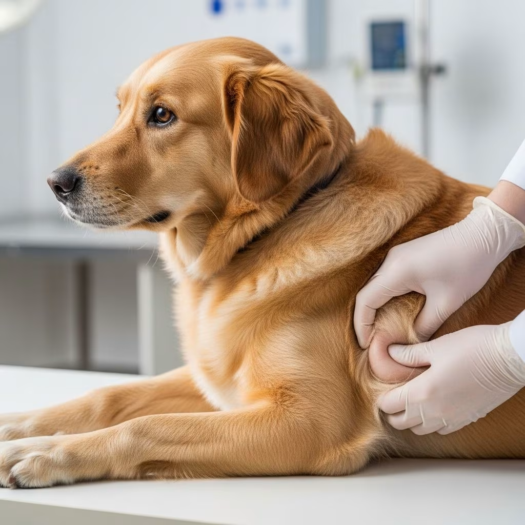
Hernias represent a significant category of structural abnormalities in veterinary medicine, where an organ or tissue protrudes through a weakened area in the surrounding muscle or connective tissue wall. These conditions can be congenital, arising from developmental flaws during fetal growth, or acquired due to trauma, increased abdominal pressure, or degenerative changes. In animals, hernias commonly affect the abdominal wall, diaphragm, and pelvic regions, leading to potential complications like organ strangulation, respiratory distress, or digestive obstruction if untreated. Early recognition and intervention are crucial for favorable outcomes, particularly in high-risk species such as dogs, horses, cattle, and sheep.
Understanding the Pathophysiology of Hernias
The development of a hernia involves a defect in the integrity of a body wall, allowing contents like intestines, fat, or even bladder to herniate. Congenital hernias stem from incomplete closure of embryonic structures, while acquired ones often result from factors like obesity, pregnancy, heavy lifting in working animals, or chronic coughing. The hernia sac, formed by peritoneum or other serosal layers, may or may not communicate freely with the peritoneal cavity. Complications arise when the hernial contents become incarcerated or strangulated, compromising blood supply and leading to necrosis—a veterinary emergency requiring immediate surgery.
In livestock, genetic predispositions play a notable role, with certain breeds showing higher incidences due to selective breeding for traits like large body size or rapid growth. Companion animals, particularly brachycephalic breeds, face risks from anatomical quirks that strain abdominal musculature.
Common Types of Hernias Observed in Veterinary Practice
Veterinarians encounter a variety of hernia types, each with distinct anatomical locations and clinical implications. Below is a breakdown of the most prevalent forms across species.
Umbilical Hernias: Frequent in Neonates
Umbilical hernias occur at the site of the umbilical cord remnant, where the abdominal wall fails to fully close post-birth. These are among the most common congenital defects in foals, calves, and puppies, often presenting as a soft, reducible swelling at the navel. While many small hernias (umbilical hernias) resolve spontaneously by 6 months as the animal grows, larger ones risk intestinal entrapment.
- Predisposed species: Foals (second most common congenital defect), calves, lambs, piglets, puppies.
- Symptoms: Visible bulge, pain on palpation if complicated, occasional vomiting or colic.
- Management: Observation for small defects; herniorrhaphy for persistent or symptomatic cases.
Inguinal Hernias: Risks in Males and Pregnant Females
Inguinal hernias involve protrusion through the inguinal canal, more common in males due to the descent of testes. In females, pregnancy exacerbates the risk. These can be direct (through weakened fascia) or indirect (via patent processus vaginalis). Horses and pigs show notable incidences, with potential for severe complications like bowel obstruction.
| Type | Species Affected | Key Risks |
|---|---|---|
| Indirect | Horses, pigs, dogs | Testicular involvement, fertility issues |
| Direct | Cattle, pregnant sows | Strangulation during farrowing |
Diaphragmatic Hernias: Life-Threatening Thoracic Protrusions
Diaphragmatic hernias feature abdominal organs entering the thoracic cavity through a diaphragmatic rent, often congenital (e.g., via the foramen of Bochdalek) or traumatic. This leads to pulmonary hypoplasia in neonates and acute respiratory distress post-trauma in adults. Common in cats (road accidents) and calves.
- Congenital form: More prevalent in brachycephalic dogs and certain cattle breeds.
- Traumatic form: History of vehicular impact or falls in large animals.
- Clinical signs: Dyspnea, cyanosis, muffled heart sounds, post-prandial distress.
Perineal and Ventral Abdominal Hernias
Perineal hernias, typical in aged intact male dogs, involve pelvic diaphragm weakness, allowing rectum, prostate, or bladder protrusion. Ventral hernias, including scrotal and femoral types, arise from midline or flank wall defects. Sheep and goats may develop ventral hernias from nutritional deficiencies or parasites weakening tissues.
Species-Specific Considerations and Breed Predispositions
Hernia prevalence varies by species, influenced by anatomy, management, and genetics.
Companion Animals: Dogs and Cats
In dogs, umbilical hernias affect breeds like English Bulldogs and Pekinese, while inguinal ones are seen in Boston Terriers. Perineal hernias plague older German Shepherds and Collie breeds due to pelvic muscle atrophy. Cats frequently suffer traumatic diaphragmatic hernias, with congenital forms rarer but reported in Siamese lines.
Large Animals: Horses, Cattle, and Small Ruminants
Foals exhibit high rates of umbilical hernias, potentially linked to in utero factors. Horses also face inguinal risks during strenuous exercise. Cattle show scrotal hernias in bulls, and sheep experience abomasal-related complications mimicking hernias. Genetic screening in breeding stock is advised for herds with recurrent cases.
Exotic and Production Animals: Pigs and Poultry
Pigs, especially inguinal hernia-prone Landrace breeds, suffer economic losses from culling affected boars. Poultry rarely develop true hernias but show prolapse mimicking symptoms.
Diagnostic Approaches for Accurate Identification
Diagnosis combines history, physical exam, and imaging. Palpation reveals reducibility and contents. Radiography with contrast (e.g., barium) visualizes herniated loops; ultrasound assesses vascularity. Advanced cases may require CT or exploratory laparotomy.
- History clues: Trauma, breed, age at onset.
- Imaging: Positive contrast for GI tract; negative for thoracic assessment.
- Differentials: Tumors, abscesses, hematomas.
Treatment Strategies: Conservative to Surgical
Small, asymptomatic hernias may be monitored, but most require surgical correction—herniorrhaphy with mesh reinforcement for large defects. Pre-op stabilization addresses dehydration, pain, and infection. Prognosis excels for uncomplicated cases (90%+ success), but strangulation drops it to 50-70%.
Post-operative care includes activity restriction, wound monitoring, and anti-inflammatories. In breeding animals, neutering prevents recurrence in perineal types.
Prevention and Genetic Management
Selective breeding avoids carrier lines; routine umbilical inspections in neonates aid early intervention. Nutritional optimization strengthens abdominal walls in growing animals. Trauma prevention (e.g., fencing for livestock) reduces acquired hernias.
Prognosis and Long-Term Outcomes
Outcomes hinge on timeliness: congenital repairs in young animals yield excellent results; delayed traumatic cases risk adhesions or recurrence (10-20%). Follow-up monitors for complications like hydrocele or infertility.
Frequently Asked Questions (FAQs)
Can umbilical hernias close on their own in puppies?
Yes, many small ones (<2 cm) resolve by 4-6 months without intervention, but veterinary evaluation is recommended.
What causes perineal hernias in dogs?
Primarily pelvic muscle weakness in aged, intact males, compounded by chronic straining or obesity.
Is surgery always necessary for diaphragmatic hernias?
Yes, due to risks of organ compromise; conservative management is rarely viable long-term.
Are hernias hereditary in livestock?
Often polygenic with breed predispositions; pedigree analysis guides breeding decisions.
How do I spot a strangulated hernia?
Signs include acute pain, non-reducible swelling, fever, and toxemia—seek emergency care.
References
References
- Congenital and Inherited Disorders of the Digestive System of Dogs — MSD Veterinary Manual. 2023. https://www.msdvetmanual.com/dog-owners/digestive-disorders-of-dogs/congenital-and-inherited-disorders-of-the-digestive-system-of-dogs
- Overview of Congenital Gastrointestinal Anomalies — MSD Manuals Professional Edition. 2024. https://www.msdmanuals.com/professional/pediatrics/congenital-gastrointestinal-anomalies/overview-of-congenital-gastrointestinal-anomalies
- Congenital Malformations of the Digestive System — CDC Surveillance Manual. 2022. https://archive.cdc.gov/www_cdc_gov/ncbddd/birthdefects/surveillancemanual/chapters/chapter-4/chapter4.7.html
- Congenital Disorders of the Equine Gastrointestinal Tract — Wiley Online Library. 2021. https://onlinelibrary.wiley.com/doi/10.1002/9781119617228.ch16
- Merck Veterinary Manual: Digestive System Anomalies — Merck Veterinary Manual. 2024. https://www.merckvetmanual.com/digestive-system/congenital-and-inherited-anomalies-involving-the-digestive-system/congenital-and-inherited-anomalies-of-the-stomach-in-animals



