

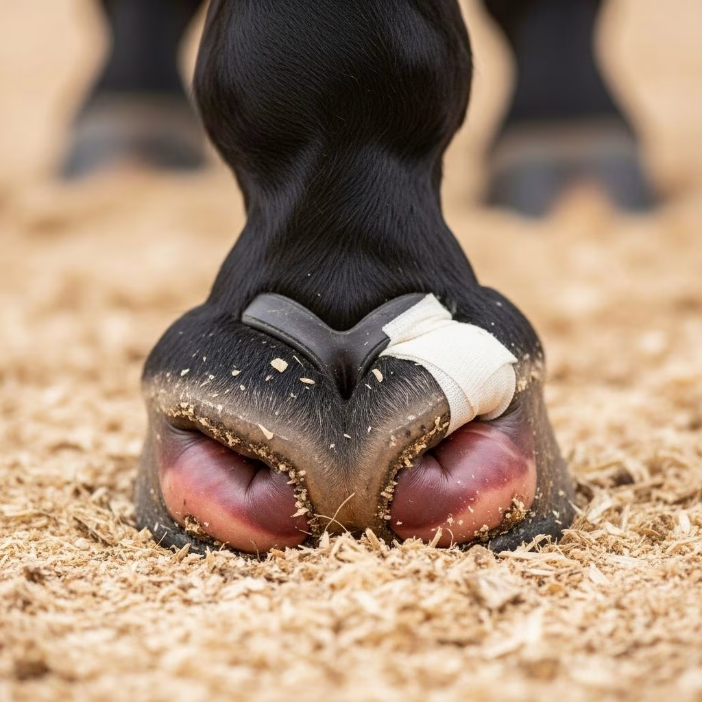
The heel bulbs represent critical soft tissue structures at the rear of a horse’s hoof, essential for shock absorption, stability, and overall foot function. Injuries to these areas, particularly lacerations, are among the most frequent traumatic wounds encountered in equine practice, often resulting from everyday activities or environmental hazards. Understanding their anatomy, vulnerability, and management is vital for horse owners, trainers, and veterinarians to facilitate swift recovery and minimize long-term complications.
Understanding Equine Hoof Anatomy: Focus on Heel Structures
The equine hoof is a marvel of biomechanical engineering, designed to withstand immense forces while providing flexibility. At the back of the hoof lies the **heel bulb**, a pair of rounded, cushioned pads composed of soft, elastic tissue that caps the digital cushion. These bulbs allow the hoof to expand and contract during movement, aiding in impact absorption and promoting blood circulation through the leg.
Internally, the heel bulbs connect to the **digital cushion**, a fibro-fatty mass that sits above the frog and beneath the pedal bone (also known as the coffin bone). This cushion transfers compressive forces from the ground upward, distributing weight evenly across the foot. Adjacent structures include the **heel buttress**, which forms a stable landing platform, and the **bars**, ridges that reinforce the heel area for added strength.
- Key Components: Heel bulbs enclose the digital cushion, interfacing with the coronary cushion for nourishment and flexibility.
- Function: Shock absorption, heel expansion, and support for the navicular bone and deep digital flexor tendon.
- Vascular Supply: Rich network including the palmar/plantar digital artery and vein, critical for healing but prone to severe bleeding if damaged.
The heel region’s mobility—essential for natural gait—makes it susceptible to repeated stress, exacerbating injuries if not properly managed. The pedal bone, the hoof’s primary weight-bearing structure, relies on these soft tissues for alignment and health. Disruptions here can lead to lameness, altered hoof growth, or chronic issues like navicular syndrome.
Common Mechanisms of Heel Bulb Trauma
Heel bulb lacerations typically arise from mechanical trauma during routine activities. Horses frequently injure themselves by **overreaching**, where the hind hoof clips the front heel bulb due to long strides, especially in breeds with exaggerated movement like Thoroughbreds or during fatigue.
Other prevalent causes include:
- Kicking Incidents: Striking barn walls, fences, or sheet metal, often resulting in deep, jagged wounds.
- Stepping on Self or Others: Common in crowded pastures or during play, leading to puncture or avulsion injuries.
- Environmental Hazards: Sharp rocks, debris, or poorly maintained footing in arenas and stalls.
- Improper Trimming: Excessive heel reduction can destabilize the foot, increasing injury risk.
These events are more common in hindlimbs due to longer stride lengths, with bell boots often recommended prophylactically. Severity varies from superficial skin tears to penetration of deeper structures like tendons, joints, or neurovascular bundles.
Assessing the Extent of Heel Bulb Lacerations
Upon noticing lameness, swelling, or bleeding at the heel, immediate veterinary evaluation is crucial. Clinical signs include:
| Symptom | Indication | Severity Level |
|---|---|---|
| Localized swelling and heat | Skin/soft tissue involvement | Mild |
| Profuse bleeding, pulsing | Digital artery laceration | Moderate-Severe |
| Lameness with toe drag | Nerve/tendon damage | Severe |
| Joint effusion, crepitus | Synovial structure penetration | Critical |
| Hoof wall deformity | Coronary band involvement | Long-term |
Diagnostic tools such as ultrasound reveal tendon integrity, while digital radiography assesses bone fragments or gas pockets indicative of infection. The neurovascular bundle’s proximity heightens risks: arterial severance causes hypovolemia, while nerve damage mimics neurectomy, leading to temporary analgesia but proprioceptive loss. Synovial involvement (e.g., digital sheath, coffin joint, navicular bursa) demands aggressive intervention to prevent septic arthritis.
Immediate First Aid for Heel Wounds
While awaiting professional care, stabilize the horse in a clean stall with bedding to minimize contamination. Key steps:
- Clean Gently: Irrigate with copious saline or dilute antiseptic; avoid high-pressure hosing on deep wounds.
- Control Hemorrhage: Apply direct pressure with sterile gauze; tourniquets are rarely needed due to digital artery size.
- Bandage Securely: Use non-adherent pads, cast padding, and vet-wrap, extending above the fetlock for compression.
- Administer NSAIDs: Phenylbutazone (2-4 mg/kg IV/PO) for pain and inflammation, per veterinary direction.
Avoid walking the horse excessively, as heel movement promotes granulation and deformity.
Advanced Treatment Strategies
Veterinary management tailors to wound depth. Superficial lacerations heal with debridement, lavage, and bandaging changes every 3-5 days, often resolving in 2-4 weeks. Deeper injuries require:
- Surgical Exploration: Under sedation/analgesia, probe for foreign bodies and repair vessels/tendons if feasible.
- Antimicrobial Therapy: Broad-spectrum IV antibiotics (e.g., penicillin + gentamicin) for 5-7 days, transitioning to oral.
- Hoof Immobilization: Bar shoes, hoof casts, or phalangeal casts prevent motion, reducing proud flesh formation.
- Regional Perfusion: For avascular risks, deliver antibiotics directly to the foot via digital artery.
Tendon/ligament damage, with poor vascularity, may necessitate tenotomy or supportive splinting. Coronary band injuries demand precise suturing to avert hoof flares or cracks.
Recovery Timeline and Monitoring
Healing varies: mild cases return to light work in 4-6 weeks; severe ones may require 3-6 months rest. Monitor for complications like abscesses, laminitis from pain-induced weight shifts, or chronic lameness. Sequential radiographs track bone remodeling, while thermography detects inflammation hotspots.
Rehabilitation includes controlled exercise, therapeutic farriery (e.g., heel elevation pads), and gradual shoeing adjustments to restore balance. Nutritional support with biotin and omega-3s aids horn quality.
Preventing Heel Bulb Lacerations
Proactive measures significantly reduce incidence:
- Protective Gear: Rubber bell boots for hind feet, especially during turnout or exercise.
- Farrier Maintenance: Regular trims balancing heels and toes to prevent overreaching.
- Stall Safety: Remove protrusions, use padded walls, and provide deep bedding.
- Footing Management: Even, cushioned surfaces in arenas; inspect pastures for debris.
- Breed-Specific: Monitor high-striding horses like Warmbloods closely.
Early detection through daily hoof checks fosters prompt intervention.
Frequently Asked Questions (FAQs)
What is the fastest way to heal a heel bulb laceration?
Prompt cleaning, bandaging, and immobilization with veterinary oversight yield the quickest recovery, often 2-4 weeks for superficial wounds.
Can heel bulb injuries cause permanent lameness?
Yes, if synovial structures or major tendons are involved without treatment, but most heal fully with proper care.
Are bell boots effective for all horses?
They excel for overreachers but fit loosely to avoid rubs; consult a farrier for sizing.
How do I know if the digital artery is cut?
Look for bright red, pulsing blood; apply pressure and call a vet immediately.
Is casting always necessary?
No, for minor wounds bandaging suffices; casts are for deep injuries needing stability.
References
- Hoof Anatomy — British Horse Society. Accessed 2026. https://www.bhs.org.uk/horse-care-and-welfare/health-care-management/hoof-care/hoof-anatomy/
- Functional Anatomy of the Equine Hoof — Scoot Boots. 2023-10-15. https://content.scootboots.com/blogs/blog/functional-anatomy-of-the-equine-hoof
- Heel Bulb Lacerations — University of Minnesota (open.lib.umn.edu). 2022-05-12. https://open.lib.umn.edu/largeanimalsurgery/chapter/heel-bulb-lacerations/
- A Deep Dive Into Horse Hoof And Foot Anatomy — Equi-Spa. 2020-05-18. https://equispa.com/2020/05/18/a-deep-dive-into-horse-hoof-and-foot-anatomy/
- Hoof Anatomy, Care and Management in Livestock (ID-321-W) — Purdue University Extension (.edu). 2018-07. https://www.extension.purdue.edu/extmedia/id/id-321-w.pdf
- Anatomy and Physiology Part 5: The Equine Foot — The Horse. 2019-11-01. https://thehorse.com/129575/anatomy-and-physiology-part-5-the-equine-foot/



