

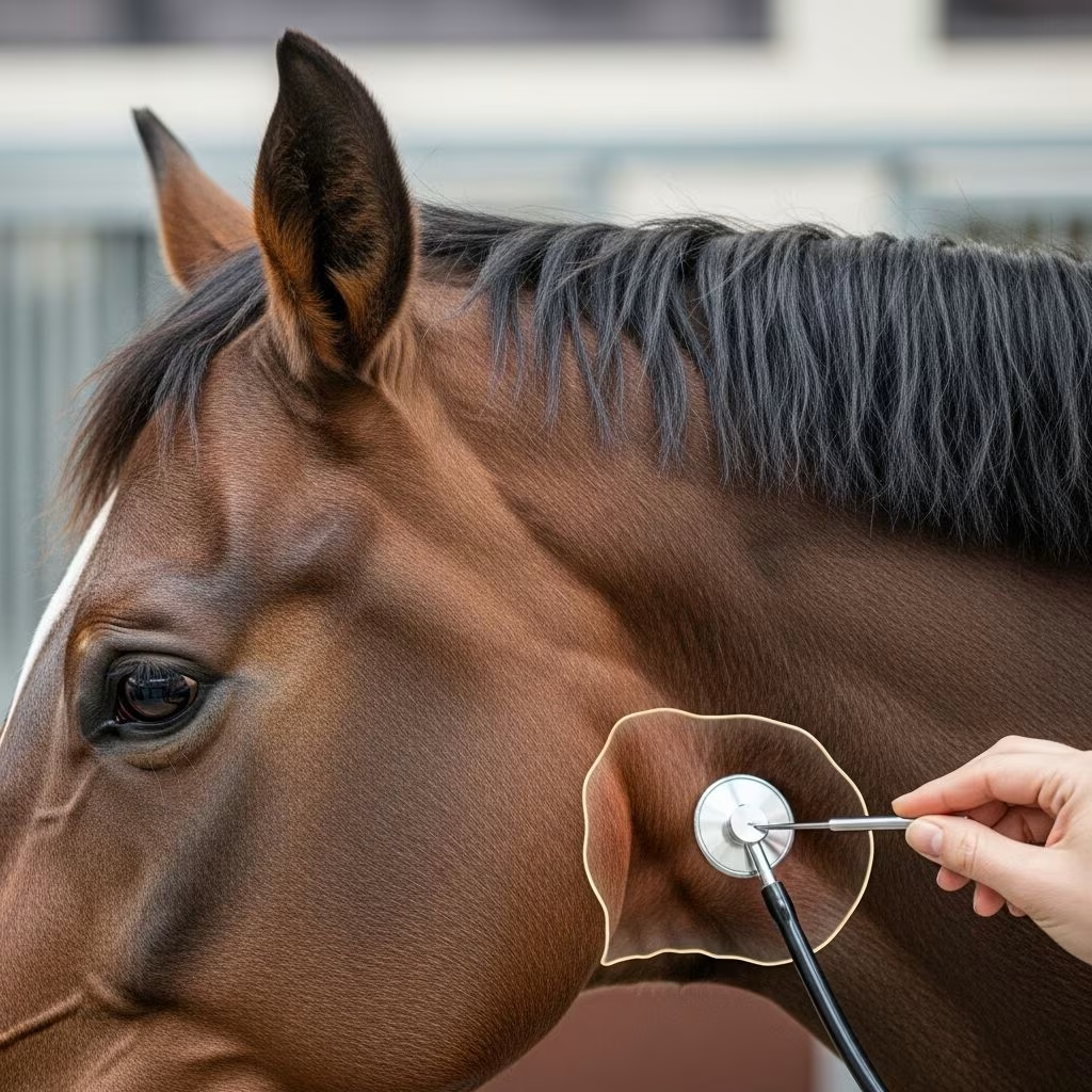
Horses possess unique anatomical features known as guttural pouches, which are spacious, air-filled sacs connected to the Eustachian tubes. These structures, located behind the jaw and within the skull, play roles in regulating ear pressure, aiding thermoregulation, and supporting swallowing. However, they are prone to various disorders that can severely impact a horse’s health, performance, and even life. Common conditions include bacterial accumulations known as empyema, fungal invasions termed mycosis, and air trapping called tympany. Early recognition of signs like unusual nasal discharge or facial swelling is crucial for effective intervention.
The Anatomy and Function of Guttural Pouches
Each horse has a pair of guttural pouches, one on either side of the throatlatch region, capable of holding up to two liters of air when fully expanded. They extend from the pharynx up toward the base of the skull, adjacent to critical structures such as major arteries, cranial nerves, and the larynx. This proximity makes infections or inflammations particularly dangerous, as they can erode vessel walls or impair nerve function.
- Primary functions: Equalizing pressure between the middle ear and atmosphere, cooling arterial blood to the brain, and assisting in swallowing by opening during deglutition.
- Vulnerable location: Shares walls with the internal carotid artery, glossopharyngeal nerve, and vagus nerve, explaining severe complications from diseases.
Understanding this anatomy helps explain why seemingly minor infections can lead to catastrophic outcomes like hemorrhage or neurological deficits.
Recognizing Bacterial Empyema: Pus Accumulation in the Pouches
Guttural pouch empyema occurs when pus from bacterial infections builds up within these sacs, often following strangles caused by Streptococcus equi. Pus may drain from ruptured lymph node abscesses into the pouches, where it stagnates and fosters ongoing infection. In roughly 20% of cases, the pus dries into hard chondroids—stone-like formations that resist flushing and serve as bacterial reservoirs, prolonging contagion risks.135
Owners might notice intermittent thick, creamy discharge from one nostril, especially after exercise or head lowering. Retropharyngeal lymph node swelling causes painful parotid region enlargement, prompting reluctance to eat or fever in acute phases. Advanced cases involve dysphagia (trouble swallowing), stertorous respiration, or cranial nerve damage manifesting as facial asymmetry or roaring during exertion.
| Symptom | Frequency | Implication |
|---|---|---|
| Unilateral purulent nasal discharge | Common | Indicates pus drainage |
| Throatlatcheswelling (painful) | Common | Lymphadenopathy |
| Fever/depression | Variable | Systemic involvement |
| Dysphagia/neuropathy | Severe cases | Nerve compression |
These signs demand prompt veterinary attention to curb spread and complications.
Fungal Mycosis: A Threatening Invasive Infection
Guttural pouch mycosis involves fungal overgrowth, predominantly Aspergillus species, thriving in the moist pouch environment, particularly in stabled mature horses inhaling spores from bedding or feed. Fungal plaques invade the mucosal lining, eroding into adjacent arteries and nerves. The hallmark is sudden, profuse epistaxis (nosebleed) from internal carotid artery rupture, which can be fatal if not ligated swiftly.125
Beyond bleeding, clinical presentations include dysphagia from glossopharyngeal or vagus nerve damage, head tilting, facial paralysis (drooping ear, eyelid, or lip), and respiratory stridor. Fluid accumulation leads to non-painful ventral throat swelling. Diagnosis hinges on endoscopic visualization of yellowish plaques and vascular changes.
- High-risk signal: Unexplained nasal hemorrhage—treat as emergency.
- Neurological signs: Indicate poor prognosis due to irreversible nerve injury.
This condition’s lethality underscores the need for vigilant monitoring in performance horses.
Guttural Pouch Tympany: Air Trapping in Young Horses
Primarily affecting foals and weanlings (birth to one year), especially fillies, tympany results from a dysfunctional pharyngeal orifice acting as a one-way valve. Excess tissue or inflammation traps air entering during swallowing, distending the pouch without pus or fungus involvement. Visible as a firm, non-painful parotid swelling, it compresses the pharynx, causing dyspnea, stridor, or aspiration pneumonia if feed enters airways.127
Unlike infections, systemic signs like fever are absent. Endoscopy reveals a ballooned pouch and ostial narrowing. While less dire, unresolved cases stunt growth or lead to secondary infections.
Diagnostic Approaches for Accurate Identification
Veterinarians employ a multi-modal strategy to pinpoint guttural pouch issues. Initial physical exams assess vital signs, palpate for swelling, and auscultate airway sounds. Endoscopy via a 1-2 meter flexible scope through the nostril offers direct visualization—gold standard for confirming pus, plaques, chondroids, or tympanic distension.147
Ultrasonography detects abscesses or fluid pockets externally, while radiography or CT scans map deeper involvement, especially pre-surgical. Hematology may reveal leukocytosis in bacterial cases, and culture/biopsy identifies pathogens for targeted therapy. Differential diagnoses include sinusitis or ethmoid hematomas.
Treatment Strategies Tailored to Condition Type
Management varies by disorder but prioritizes lavage, antimicrobials, and surgery.
For Empyema
Repeated saline flushes via indwelling catheter remove pus and chondroids (manual extraction or laser fragmentation if needed). Systemic antibiotics (penicillins for streptococci) last 4-6 weeks, with topical antiseptics preventing recurrence. Urovaxin vaccination reduces strangles-related risks.
For Mycosis
Urgent arterial ligation (surgical or balloon catheterization) halts bleeding, followed by antifungal washes (imidazoles) and systemic therapy. Plaque debridement via endoscopy aids resolution, though nerve damage persists.
For Tympany
Foil puncture temporarily relieves pressure; definitive fix is surgical ostial enlargement (plicaectomy) under general anesthesia, yielding excellent outcomes.345
Supportive care includes NSAIDs for pain, hydration, and isolation for contagious cases.
Prevention Measures for Horse Owners
Proactive steps mitigate risks: vaccinate against strangles, ensure ventilation in stalls to minimize fungal spores, and quarantine new arrivals. Routine endoscopy for racing horses detects subclinical issues. Nutritional balance supports immunity, and prompt upper respiratory treatment averts progression to pouches.
- Monitor foals for early swelling.
- Avoid dusty hay for housed adults.
- Annual vet checks for high-value equines.
Prognosis and Long-Term Outlook
Empyema carries good prognosis with aggressive lavage (90% resolution), though chondroids complicate. Mycosis survival hinges on bleed control (50-70% if addressed early), with dysphagia signaling guarded outlook. Tympany responds best to surgery (>95% success). Recurrence is low with adherence, but performance horses may suffer chronic roaring.15
Frequently Asked Questions (FAQs)
What triggers guttural pouch empyema most often?
Bacterial spread from strangles or upper airway infections, leading to pus entrapment.
Is nosebleeding always fatal in mycosis?
No, but it requires immediate surgery; delays increase mortality.
Can tympany resolve without treatment?
Rarely; persistent cases risk pneumonia or growth issues.
How much does treatment cost?
Varies: endoscopy $500-1000, surgery $3000-8000, plus meds.
Are guttural pouches unique to horses?
Yes, exclusive to Equidae, absent in other equines like donkeys partially.
Key Takeaways for Equine Care
- Guttural pouch health is pivotal for airway integrity.
- Observe for discharge, swellings, or bleeds daily.
- Endoscopy is diagnostic cornerstone.
- Tailored treatments yield high success rates.
References
- Guttural Pouch Disease in Horses — Merck Veterinary Manual. 2023. https://www.merckvetmanual.com/horse-owners/lung-and-airway-disorders-of-horses/guttural-pouch-disease-in-horses
- Guttural Pouch Disease in Horses – Respiratory System — MSD Veterinary Manual. 2023. https://www.msdvetmanual.com/respiratory-system/respiratory-diseases-of-horses/guttural-pouch-disease-in-horses
- Equine Guttural Pouch Infections — The Horse. 2018-10-01. https://thehorse.com/194583/equine-guttural-pouch-infections/
- Guttural Pouch Disease in Horses – Causes, Treatment — Vetster. 2023. https://vetster.com/en/conditions/horse/guttural-pouch-disease
- Guttural Pouch Disease in Horses: Causes, Treatment, & Prognosis — Mad Barn. 2023. https://madbarn.com/guttural-pouch-disease-in-horses/
- Diagnostic Imaging of Diseases Affecting the Guttural Pouch — PMC (PubMed Central). 2023-08-29. https://pmc.ncbi.nlm.nih.gov/articles/PMC10458280/



