

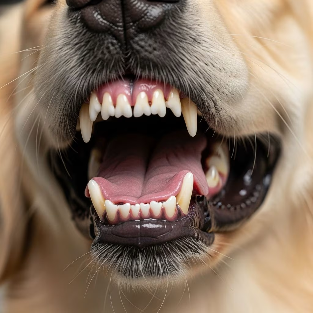
Periodontal disease stands as the leading oral health concern for dogs and cats, impacting the structures supporting their teeth and potentially leading to severe complications if ignored. This condition arises from bacterial buildup that triggers inflammation, affecting gums, bone, and ligaments around the teeth.
Understanding the Foundations of Pet Oral Pathology
The oral cavity in small animals hosts a complex ecosystem where bacteria thrive on food particles and saliva, forming a sticky layer known as plaque. Within hours, this plaque mineralizes into hard tartar, irritating the gums and initiating gingivitis, the earliest reversible stage. Progression to periodontitis involves deeper tissue destruction, including bone resorption, which compromises tooth stability.
Key anatomical components include the gingiva (gums), periodontal ligament, cementum covering roots, and alveolar bone. Disruption here not only causes local pain but can disseminate bacteria systemically, linking to heart, liver, and kidney issues.
Prevalence and Vulnerable Populations
Statistics reveal alarming rates: by age 2, approximately 80% of dogs and 70% of cats exhibit signs of periodontal involvement, escalating with age and size. Smaller breeds like Chihuahuas, Yorkshire Terriers, and Dachshunds face heightened risks due to crowded teeth, minimal bone support, and plaque-trapping anatomy. Brachycephalic dogs (e.g., Pugs, Boxers) suffer from malocclusion, exacerbating buildup.
Systemic factors amplify susceptibility: diabetes, Cushing’s disease, and immunosuppression weaken defenses, while poor diet or infrequent grooming accelerates onset. Cats show correlations with body weight increases and age.
| Factor | Dogs Affected | Cats Affected |
|---|---|---|
| By Age 2 | 80% | 70% |
| Small/Toy Breeds | High Risk | Moderate |
| Age >3 Years | 80-90% | High |
Recognizing Early Warning Signals
Owners often overlook subtle cues, delaying intervention until advanced stages. Common indicators include persistent foul breath (halitosis), visible yellow-brown tartar, red or bleeding gums during play or eating, and drooling. Advanced signs encompass tooth mobility, facial swelling, pawing at the mouth, appetite loss, and favoring one side.
- Halitosis: First and most prevalent sign from bacterial overgrowth.
- Gingival Inflammation: Swollen, receding gums exposing roots.
- Pocketing: Gaps between teeth and gums harboring debris.
- Bone Loss: Detected via imaging, leading to furcation exposure on multi-rooted teeth.
In cats, similar patterns emerge, though they mask pain adeptly, manifesting as reduced grooming or litter avoidance.
Diagnostic Approaches for Accurate Assessment
Effective management begins with thorough evaluation under general anesthesia, as conscious exams miss subgingival pathology. Initial steps involve visual inspection and periodontal probing to measure pocket depths—normal for dogs is 1-3 mm, cats 0.5-1 mm; excesses signal attachment loss.
Intraoral radiography is indispensable, revealing 60-70% of hidden bone loss invisible externally, per AAHA guidelines. Advanced cases may warrant CT for surgical planning.
- Pre-anesthetic oral survey.
- Anesthetized probing and charting.
- Full-mouth radiographs.
- Staging based on attachment loss percentage.
Therapeutic Interventions by Stage
Treatment scales with severity, aiming to halt progression, alleviate pain, and preserve viable dentition.
Stage 1: Gingivitis Reversal
Professional scaling removes supragingival plaque/tartar, polishing prevents recurrence. Home brushing daily with enzymatic toothpaste suffices.
Stage 2: Mild Periodontitis
Subgingival scaling/root planing and curettage target pockets <4 mm. Antimicrobials like chlorhexidine gels aid healing.
Stage 3: Moderate Attachment Loss
Open flap surgery exposes roots for debridement, possibly with guided tissue regeneration or bone grafts for infrabony defects.
Stage 4: Severe Disease
Extractions for non-salvageable teeth predominate, often 20-50% in advanced cases. Oronasal fistula repairs or endodontic therapy combine for complicated lesions. Post-op antibiotics, analgesics, and e-collars ensure recovery.
Prognosis improves with early action; late-stage salvages rarely exceed 50% success.
Empowering Owners: Home Maintenance Strategies
Professional care alone falters without daily owner commitment. Toothbrushing every 24-48 hours with pet-safe products mechanically disrupts plaque.
- Dental diets with fiber to scrape teeth.
- Chews/toys (VOHC-approved) reducing tartar by 20-70%.
- Water additives, rinses, gels with enzymes/antiseptics.
- Regular vet checks: 6-12 months for at-risk pets.
Avoid human toothpaste (xylitol toxic to dogs). Consistency yields measurable calculus reduction in weeks.
Preventive Blueprint for Lifelong Oral Wellness
Initiate care at puppy/kitten stage: weekly exams, introduce brushing early. Annual cleanings under anesthesia for adults, biannual for predisposed. Nutritional balance supports immunity; obesity control mitigates feline risks.
Monitor for comorbidities; diabetic pets require tighter regimens. Education empowers: apps tracking brush sessions boost compliance.
Systemic Ramifications and Holistic Care
Bacteremia from diseased sites seeds distant organs, elevating endocarditis, nephropathy risks—studies link severe periodontitis to 2-3x organ failure odds. Holistic vets integrate probiotics, omega-3s for anti-inflammatory effects.
Frequently Asked Questions
What breeds are most prone to gum disease?
Toy and small breeds like Yorkies, Chihuahuas, Pugs due to anatomy.
Can periodontal disease be reversed?
Gingivitis yes, via cleaning; periodontitis managed but bone loss permanent.
How often should I brush my pet’s teeth?
Daily or every other day for best results.
Is anesthesia safe for dental cleanings?
Yes, when pre-op health screened; essential for thorough treatment.
Do dental treats really work?
VOHC-stamped ones reduce plaque effectively as adjuncts.
Conclusion: Prioritizing Pet Dental Vitality
Proactive vigilance transforms periodontal disease from inevitable scourge to manageable condition, extending pain-free years. Partner with vets for tailored plans.
References
- Periodontal Disease in Small Animals — Merck Veterinary Manual. 2023. https://www.merckvetmanual.com/digestive-system/dentistry-in-small-animals/periodontal-disease-in-small-animals
- Periodontal Disease in Dogs — Small Door Veterinary. 2023. https://www.smalldoorvet.com/learning-center/medical/periodontal-disease-in-dogs
- Periodontal disease — PubMed (Cornell University). 2008-05-01. https://pubmed.ncbi.nlm.nih.gov/18482707/
- Periodontal Disease in Dogs and Cats — Today’s Veterinary Nurse. 2023. https://todaysveterinarynurse.com/dentistry/periodontal-disease-in-dogs-and-cats/
- Periodontal Disease — Texas A&M Veterinary Medical Teaching Hospital. 2023. https://vethospital.tamu.edu/small-animal/dentistry/periodontal-disease/
- Periodontal disease — Cornell University College of Veterinary Medicine. 2023. https://www.vet.cornell.edu/departments-centers-and-institutes/riney-canine-health-center/health-topics/canine-health-information/periodontal-disease
- Periodontal Disease in Pets — AVMA. 2023. https://ebusiness.avma.org/files/productdownloads/petdentalcare_brochure.pdf



