

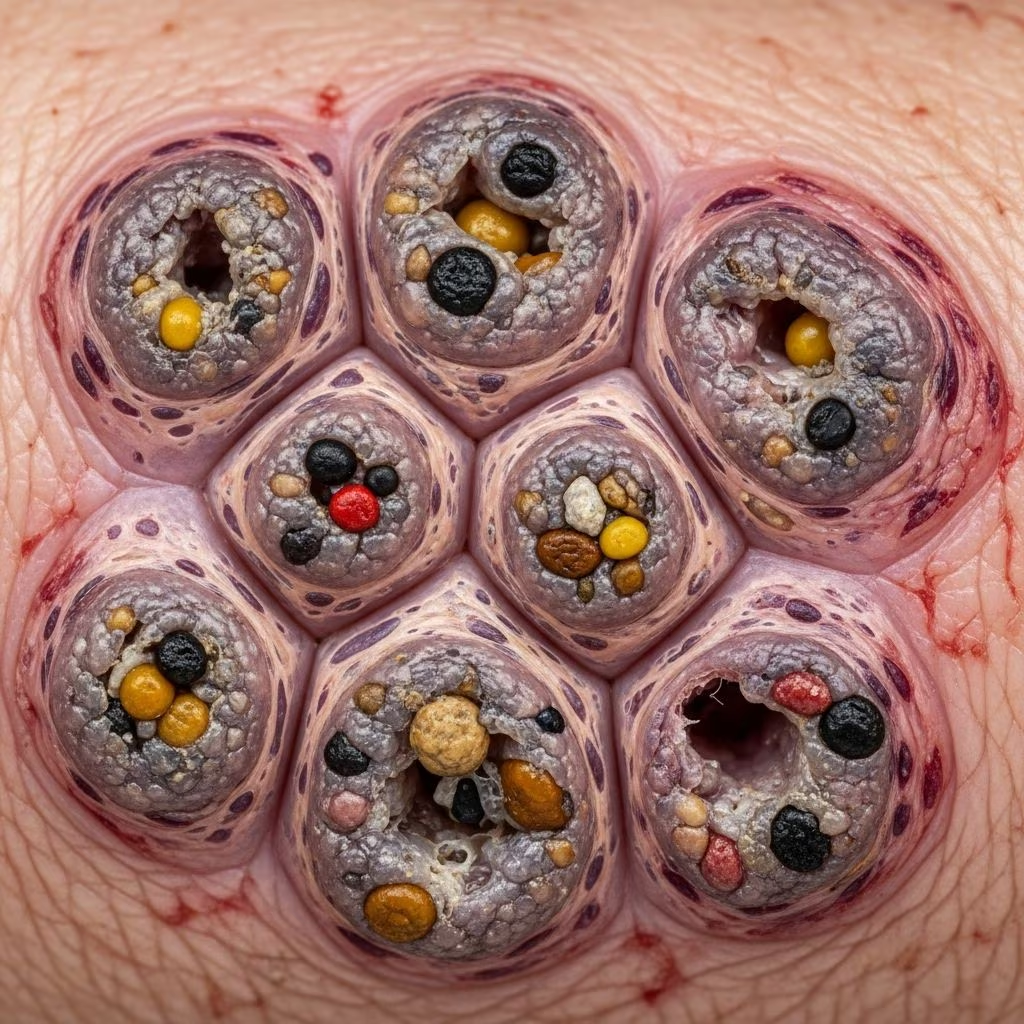
Mycetomas represent a distinctive class of chronic infections that affect the subcutaneous tissues of various animal species. These condition constitute a significant challenge in veterinary medicine due to their resistance to conventional treatment and their potential to cause substantial tissue damage if left untreated. Understanding the nature of mycetomas, their causative organisms, clinical manifestations, and available therapeutic approaches is essential for veterinarians managing affected animals.
Understanding the Nature of Mycetomas
A mycetoma is fundamentally a granulomatous nodular lesion that develops within the subcutaneous tissue layers beneath the skin. The hallmark characteristic of mycetomas is the formation of visible grain-like structures composed of tightly aggregated fungal organisms suspended within a hard, cement-like matrix. These grains, which can range in color from black to white depending on the causative fungal species, give mycetomas their distinctive appearance and are crucial for diagnosis.
The disease develops through a chronic inflammatory response wherein the animal’s immune system attempts to wall off the fungal invaders through the formation of pyogranulomatous tissue. This inflammatory process, while protective, simultaneously contributes to progressive tissue destruction and the characteristic draining tracts observed in advanced cases. The infection typically progresses slowly, with affected animals sometimes showing minimal systemic signs despite significant local tissue involvement.
Classification and Causative Organisms
Mycetomas in animals are categorized based on the type of causative organism. Eumycotic mycetomas, caused by fungi, represent the most commonly encountered form in veterinary practice, though bacterial-induced mycetomas also occur. Within the fungal category, organisms are further differentiated by the color of the grains they produce:
- Black-grain eumycetomas: Typically caused by dematiaceous (pigmented) fungi including Curvularia species, Madurella species, Cladophialophora bantiana, and Pseudallescheria boydii
- White-grain mycetomas: Often associated with non-pigmented fungal species and sometimes bacterial organisms
The geographic distribution of mycetoma-causing fungi varies significantly. Madurella species predominate in tropical and subtropical regions such as India, Africa, and South America, where they remain endemic. In temperate climates, mycetomas are considerably rarer, though sporadic cases have been documented in Europe and other non-tropical areas, often in animals with outdoor exposure to contaminated soil.
Pathophysiology and Transmission
Mycetomas develop exclusively through traumatic implantation of fungal spores directly into subcutaneous tissues. Unlike respiratory fungal infections that may be acquired through inhalation, mycetomas require a breach in skin integrity through which environmental spores gain entry into deeper tissue layers. Common routes of infection include puncture wounds from thorns, grass awns, contaminated soil exposure, or other traumatic injuries that animals may incur while outdoors.
Once implanted, the fungal organisms establish a localized chronic infection. The infection typically remains confined to the inoculation site and surrounding tissues, though in some cases, particularly with abdominal mycetomas, the infection may extend more extensively through tissue planes. The slow progressive nature of mycetomas reflects the organism’s growth rate and the ongoing inflammatory response mounted by the host immune system.
Clinical Presentation and Affected Sites
The clinical manifestations of mycetomas vary depending on their anatomical location and the extent of tissue involvement. Cutaneous and subcutaneous mycetomas represent the most frequently encountered presentations:
- Subcutaneous nodules or swelling on the extremities, particularly affecting the legs and feet
- Facial involvement with visible swelling on the muzzle or bridge of the nose
- Draining tracts that discharge fluid containing characteristic grains
- Progressive enlargement of the affected area over weeks to months
Black-grain mycetomas demonstrate a distinct predilection for the appendicular skeleton and facial regions. When mycetomas involve the feet or limbs, the infection may progress to involve underlying bone structures, potentially complicating treatment significantly. Some animals develop nodules on the head and neck, while others present with lesions on the thoracic wall or chest region.
Abdominal mycetomas constitute a more serious presentation, potentially involving visceral structures and peritoneal tissues. These cases often present with masses within the abdominal cavity and inflammation of the peritoneal membranes lining the abdominal walls, frequently discovered incidentally during surgery or imaging for other conditions.
Despite the chronic nature and progressive tissue involvement characteristic of mycetomas, affected animals typically experience minimal systemic signs. Fever, lethargy, and appetite changes are uncommon unless the infection has disseminated extensively. This disparity between the severity of local tissue damage and the absence of systemic illness can delay diagnosis, as owners may not recognize the gradual enlargement as a serious medical condition.
Diagnostic Approaches
Accurate diagnosis of mycetomas relies on identification of the characteristic grains within infected tissue or exudate. Several diagnostic modalities contribute to establishing a definitive diagnosis:
Grain Identification
The most direct diagnostic approach involves grain examination from exudate or tissue samples. Grains should be carefully expressed from draining wounds or removed during tissue biopsy, then palpated between fingers to assess their consistency and character. Microscopic examination of grains reveals the presence of fungal elements, with higher magnification allowing differentiation between fungal hyphae and associated inflammatory material.
Cytological and Histological Assessment
Fine needle aspiration cytology can reveal numerous black or colored foci of amorphous material surrounded by inflammatory cells including neutrophils, epithelioid macrophages, multinucleated giant cells, and lymphocytes. Histological examination of tissue biopsies demonstrates characteristic pyogranulomatous panniculitis with intralesional pigmented fungi. Special staining techniques such as Periodic Acid-Schiff (PAS) and Grocott stains enhance visualization of fungal elements within tissue sections. The Splendor-Hoeppli phenomenon—radially arranged eosinophilic material surrounding fungal aggregates—provides additional histological confirmation.
Culture and Molecular Identification
Fungal culture remains the gold standard for organism identification, though culture results may take weeks to months. Molecular techniques including 28S LSU rRNA amplification and sequencing provide rapid and accurate species identification, even from formalin-fixed paraffin-embedded tissue samples. Molecular phylogeny has proven particularly valuable in identifying emerging pathogenic species and expanding understanding of mycetoma epidemiology.
Imaging Studies
Radiographic and advanced imaging studies help assess the extent of tissue involvement, detect osseous involvement, and evaluate for abdominal lesions. These imaging modalities guide surgical planning and treatment decisions but cannot definitively diagnose mycetomas without tissue confirmation.
Treatment Strategies and Therapeutic Outcomes
Management of mycetomas presents substantial therapeutic challenges, with treatment success varying significantly based on disease extent, location, and organism type. Available treatment approaches include surgical intervention and antifungal chemotherapy, often used in combination.
Surgical Management
Surgical removal of affected tissue represents the most potentially definitive treatment approach for localized cutaneous mycetomas. Complete excision of the lesion with adequate margins of normal tissue offers the best chance for resolution. However, complete surgical removal is not always feasible, particularly when the lesion is extensive or involves critical structures. In some cases, particularly when cutaneous mycetomas involve the limbs extensively, amputation may become the only effective surgical option.
Antifungal Chemotherapy
Antifungal medication efficacy has been reported in only a limited number of mycetoma cases, though some successes have been documented. Itraconazole represents one of the more commonly employed agents, administered at doses ranging from 5-10 mg/kg once daily. Treatment protocols may employ pulse therapy—alternating weeks of treatment with treatment-free intervals—to manage cost and potential adverse effects while maintaining therapeutic effect. Itraconazole therapy may continue for extended periods, with some cases requiring treatment for 45 days or longer before lesions become clinically unappreciable.
Fluconazole has demonstrated success in isolated cases, with one documented report of intra-abdominal maduromycosis resolution using 50 mg every 24 hours for 6 weeks. However, the inconsistent efficacy of antifungal agents across different cases and organisms makes predicting treatment success challenging.
Treatment Prognosis
The prognosis for mycetoma resolution varies substantially based on multiple factors. Cutaneous mycetomas, while non-life-threatening, often prove exceptionally difficult to resolve completely. Abdominal mycetomas carry a notably guarded prognosis due to the extensive tissue involvement typical of intra-abdominal disease. Success rates improve when surgical removal can be performed completely, particularly for localized skin lesions detected early in disease progression.
Epidemiological Significance and Public Health Considerations
While mycetomas primarily affect animals, the detection of fungal mycetomas in domestic animals holds important epidemiological implications for human health. Animals living in endemic regions or with exposure to contaminated soil serve as sentinels for the presence of mycetoma-causing fungi in specific geographic areas. The identification of Madurella pseudomycetomatis in a dog in Italy expanded understanding of this organism’s geographic distribution beyond previously recognized endemic regions.
The shared route of transmission—environmental fungal spores gaining access through traumatic skin inoculation—means that animals and humans in the same geographic region face similar infection risk. Documentation of mycetoma cases in animals may alert public health authorities to fungal species present in specific regions and prompt enhanced surveillance or preventive measures.
Prevention and Risk Mitigation
While complete prevention of mycetoma is not always possible, several measures can reduce infection risk:
- Prompt treatment of puncture wounds and skin injuries to minimize fungal spore implantation risk
- Limitation of outdoor exposure in animals with known traumatic injuries or immunosuppression
- Regular examination of outdoor-dwelling animals for new wounds or subcutaneous swellings that might indicate early mycetoma development
- Awareness of geographic risk factors, particularly for animals in tropical and subtropical regions where mycetoma-causing fungi are endemic
Current Research and Future Directions
Ongoing research continues to expand understanding of mycetoma epidemiology, causative organism identification, and treatment efficacy. Molecular identification techniques have revolutionized species recognition and enabled documentation of previously unrecognized organisms in veterinary mycetoma cases. Future therapeutic developments may involve investigation of combination antifungal regimens, improved delivery systems for antifungal agents to affected tissues, and immunomodulatory approaches to enhance host response to infection.
Conclusion
Mycetomas represent a distinctive chronic fungal infection affecting subcutaneous tissues in various animal species. These infections develop through traumatic implantation of environmental fungal spores and progress slowly, often with minimal systemic illness despite progressive local tissue damage. Diagnosis relies on identification of characteristic grains within tissue and exudate, with molecular techniques providing rapid species identification. Treatment remains challenging, with surgical removal offering the most definitive approach for localized lesions, while antifungal chemotherapy demonstrates variable efficacy. Early recognition and comprehensive management offer the best opportunity for successful resolution of these debilitating infections.
References
- Canine eumycetoma caused by Madurella pseudomycetomatis — PMC/NIH. 2022. https://pmc.ncbi.nlm.nih.gov/articles/PMC8828985/
- Fungal Infections in Dogs — Dog Owners Section — Merck Veterinary Manual. 2024. https://www.merckvetmanual.com/dog-owners/disorders-affecting-multiple-body-systems-of-dogs/fungal-infections-in-dogs
- Mycetomas in Animals — Infectious Diseases — MSD Veterinary Manual. 2024. https://www.msdvetmanual.com/infectious-diseases/fungal-infections/mycetomas-in-animals
- Approaches to Opportunistic Fungal Infections in Small Animals — Today’s Veterinary Practice. 2024. https://todaysveterinarypractice.com/internal-medicine/approaches-to-opportunistic-fungal-infections-in-small-animals/
- Clinical Overview of Mycetoma — Centers for Disease Control and Prevention. 2024. https://www.cdc.gov/mycetoma/hcp/clinical-overview/index.html
- Mycetoma in Animals: A Review of Cases Reported From 1925–2022 — Qeios Research Platform. 2022. https://www.qeios.com/read/ZLD37V



