

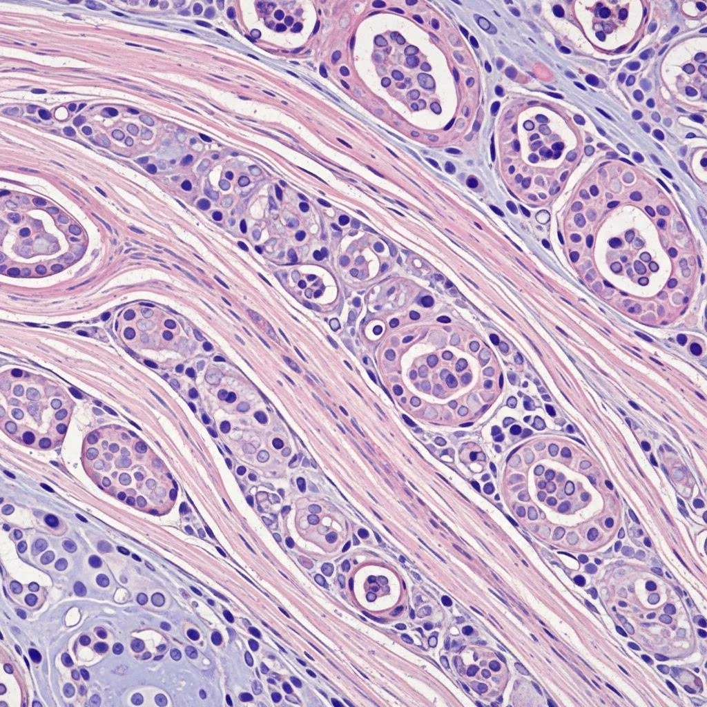
Understanding Fibrous Tumors and Hamartomas in Pets
Fibrous tumors and hamartomas are common benign skin growths that pet owners often encounter. These non-cancerous masses can appear on dogs, cats, and other animals, causing concern even though they are typically harmless. Understanding what these growths are, how they develop, and when treatment may be necessary helps pet owners make informed decisions about their companion’s health.
What Are Fibrous Tumors?
Fibrous tumors, also known as fibromas, are slow-growing, benign masses that develop from fibrous connective tissue. These tumors form as a result of chronic irritation, repeated trauma, or areas of increased pressure on the skin. Fibromas are composed primarily of mature fibrocytes and collagen fibers arranged in a dense, well-demarcated structure. Unlike malignant tumors, fibromas do not metastasize or spread to other parts of the body.
These growths typically appear as plaques, nodules, or small masses on or within the skin, commonly found in areas subject to friction or repeated mechanical stress. The development of fibrous tumors is a benign process, meaning the cells remain normal and non-cancerous throughout their growth.
Defining Hamartomas
Hamartomas represent a unique category of skin lesions that exist between developmental disorders and benign tumors. A hamartoma is defined as a focal, disorganized overgrowth of well-differentiated tissue elements that are normally present at that anatomical site. The key distinguishing feature of hamartomas is that they comprise mature tissue components arranged in an abnormal, haphazard pattern rather than following the typical architectural organization of that tissue.
Hamartomas differ fundamentally from true neoplastic processes (cancer) in that they represent disorganized proliferation of normal tissue components rather than abnormal, cancerous growth. The cells within hamartomas are phenotypically normal and fully differentiated; however, their spatial organization and proportions deviate significantly from the usual histological pattern of the affected organ.
Types of Benign Fibrous and Hamartomatous Growths
Several distinct types of fibrous tumors and hamartomas affect pets, each with characteristic features and clinical presentations:
Lipomas
Lipomas are benign tumors of adipose tissue that are perhaps more accurately characterized as hamartomas. These growths are among the most common benign tumors in dogs, occasionally identified in cats and horses, and rare in other domestic species. In dogs, lipomas generally develop in older, obese females, most commonly on the trunk and proximal limbs. The breeds at highest risk include Doberman Pinschers, Labrador Retrievers, Miniature Schnauzers, and mixed-breed dogs.
Vascular Hamartomas
Vascular hamartomas are characterized by multiple reddish papular growths that appear as proliferative vascular channels lined by a single layer of endothelial cells and supported by fibrous stroma. These lesions often demonstrate variable degrees of differentiation. Vascular hamartomas present with multiple reddish papular growths, typically ranging from 0.2 to 2 centimeters in size, frequently noticed on the ventrolateral abdomen or other areas of the body.
Follicular Hamartomas
Follicular hamartomas are considered a variant of fibroadnexal hamartoma characterized by preservation of normal follicular architecture, absence of significant atypia, and association with mature adnexal components within a fibrous matrix. These lesions typically present as round, firm masses with a white and glistening cut surface. Follicular hamartomas are distinguished from trichoepitheliomas, which often present as cystic nodules with complex follicular differentiation including multiple keratin-filled cysts.
Folliculosebaceous Hamartomas
Folliculosebaceous hamartomas are marked by proliferations of distorted follicular, sebaceous, and apocrine elements embedded within a dense fibrous stroma, generally with no cellular atypia and exhibiting a higher stromal-to-follicular ratio than simple follicular hamartomas. It is important to distinguish folliculosebaceous hamartomas from folliculosebaceous cystic hamartomas, which are defined by a central, cystically dilated folliculosebaceous structure lined with infundibular epithelium and accompanied by abundant, well-differentiated sebaceous lobules.
Epidermal Hamartomas (Nevi)
Epidermal hamartomas, also called nevi, are dark, pointy bumps on the skin that occasionally appear arranged in a line. These growths are rare and found most often in puppies. The disease may be inherited in Cocker Spaniels. Some epidermal hamartomas form pimples or dark, thick skin folds. While benign, their appearance can be unpleasant, and they are prone to secondary bacterial infection.
Epidemiology and Risk Factors
Hamartomas and fibrous tumors occur across various animal populations with distinct epidemiological patterns. Cutaneous hamartomas in dogs show relatively low incidence rates, with studies indicating approximately 5% of cutaneous masses examined being diagnosed as hamartomas. Most dogs presenting with hamartomas are mixed breeds, with a median age around 10.5 years, though hamartomas can also occur in younger animals, particularly puppies with epidermal hamartomas.
Risk factors for developing these benign growths include age, breed predisposition, obesity (particularly for lipomas), and areas of chronic irritation or repeated trauma. Older animals, particularly obese females in certain breeds, show higher incidence rates for lipomatous hamartomas.
Diagnosis of Fibrous Tumors and Hamartomas
Accurate diagnosis requires a combination of clinical evaluation and histopathological examination. Veterinarians typically perform the following diagnostic procedures:
Clinical Examination
Physical examination reveals the appearance, location, size, and characteristics of the growth. Benign fibrous tumors and hamartomas typically present as slow-growing, well-demarcated masses that do not appear to cause systemic illness or affect the animal’s overall health status.
Histopathological Examination
Definitive diagnosis requires microscopic examination of tissue samples obtained through biopsy or surgical removal. Hematoxylin and eosin (HE) staining is routinely performed, and specialized stains such as Masson’s trichrome (MT), Gomori, and Luxol fast blue may be employed to further characterize tissue composition. For accurate classification and diagnosis, MT in combination with α-SMA and neurofilament immunohistochemistry is recommended in addition to routine HE staining.
Histological Characteristics
Hamartomas are characterized by the absence of cellular atypia and a well-organized albeit haphazard tissue architecture, all indicative of a benign process. Vascular hamartomas show vascular spaces resembling normal vessels lined by normal endothelial cells. Follicular and folliculosebaceous hamartomas demonstrate normal tissue components arranged in an abnormal pattern with increased fibrous tissue supporting the epithelial components.
Treatment Options
Treatment decisions for fibrous tumors and hamartomas depend on several factors including the size, location, growth rate, and whether the lesion is causing clinical problems.
Surgical Removal
Surgical excision is the most common and definitive treatment for fibrous tumors and hamartomas. Smaller hamartomas can be surgically removed with straightforward excision techniques. For lesions with potential for recurrence or those located in sensitive areas, veterinarians may remove a wide margin of tissue surrounding the tumor to ensure complete removal and minimize recurrence risk.
Medical Management
Dogs with large or multiple hamartomas may respond to drug treatment rather than surgical intervention. Medical management options may include topical treatments to prevent secondary infection or oral medications to manage associated inflammation or irritation.
Observation
Because these tumors are benign and typically grow slowly, many cases may be managed with regular monitoring rather than immediate intervention. If the growth is not causing discomfort, secondary infection, or cosmetic concerns, observation may be appropriate.
Complications and Secondary Concerns
While fibrous tumors and hamartomas are benign, they can develop complications that necessitate treatment. Epidermal hamartomas, despite being benign, are prone to secondary bacterial infections due to their location and appearance. Secondary infections can cause discomfort and may require antibacterial treatment or surgical intervention.
Some hamartomas may grow in locations that interfere with normal movement or cause cosmetic concerns for owners. In such cases, surgical removal may be recommended for the animal’s comfort and quality of life.
Differentiation from Malignant Tumors
It is crucial to distinguish benign fibrous tumors and hamartomas from malignant tumors such as hemangiosarcomas and malignant fibrous histiocytomas. Hemangiosarcomas are malignant, exhibiting pronounced cellular atypia, increased mitotic activity, and irregular vascular structures, contrasting sharply with the benign characteristics of vascular hamartomas.
Malignant fibrous histiocytomas are rare in dogs but represent sarcomas of intermediate malignancy that are locally invasive and tend to recur after surgical removal, though they seldom metastasize. Radical excision is recommended for these lesions, with local control possibly improved by adjuvant electroporation or radiation therapy.
Prognosis and Long-term Management
The prognosis for animals with fibrous tumors and hamartomas is generally excellent. These benign lesions do not spread to other parts of the body and do not typically affect the animal’s overall health or lifespan. Most animals live normal, healthy lives with these growths present.
Long-term management involves regular monitoring of the lesion’s size and appearance. Pet owners should report any rapid growth, signs of infection, or behavioral changes related to the tumor to their veterinarian. Most benign fibrous tumors remain stable in size over many years.
Frequently Asked Questions
Q: Are fibrous tumors and hamartomas cancerous?
A: No, fibrous tumors and hamartomas are benign, non-cancerous growths. They do not spread to other parts of the body and do not represent cancer. However, it is important to have any new skin growth evaluated by a veterinarian to confirm the diagnosis.
Q: Do fibrous tumors need to be removed?
A: Not always. If the tumor is not causing discomfort, infection, or cosmetic concerns, observation may be appropriate. Surgical removal is recommended if the growth becomes infected, interferes with movement, or if the owner prefers removal for aesthetic reasons.
Q: Can hamartomas grow back after surgical removal?
A: True hamartomas rarely recur after complete surgical removal since they are developmental anomalies rather than aggressive neoplasms. However, if incomplete excision occurs, regrowth is possible.
Q: How can I tell if my pet’s skin growth is a fibrous tumor?
A: Only a veterinarian can definitively diagnose a skin growth through physical examination and biopsy. Typical characteristics of benign fibrous tumors include slow growth, well-defined margins, and lack of systemic symptoms. Bring any new or changing skin growth to your veterinarian’s attention.
Q: Are certain breeds more prone to developing these tumors?
A: Yes, certain breeds show higher predisposition to specific types of hamartomas. For example, Cocker Spaniels may inherit epidermal hamartomas, while Doberman Pinschers, Labrador Retrievers, and Miniature Schnauzers are at higher risk for lipomas.
Q: What should I do if I notice a new skin growth on my pet?
A: Schedule an appointment with your veterinarian for evaluation. They will perform a physical examination and may recommend a biopsy to determine the exact nature of the growth. Early evaluation helps ensure appropriate management and rules out any concerning conditions.
References
- Cutaneous hamartomas in dogs: Epidemiology and pathology — Veterinary Paper. 2025-01-01. https://www.veterinarypaper.com/pdf/2025/vol10issue7/PartA/10-6-38-697.pdf
- Hamartomas of the alimentary tract in dogs: A case series — SAGE Journals. 2024-01-15. https://journals.sagepub.com/doi/10.1177/03009858251345829
- Connective Tissue Tumors in Animals – Integumentary System — Merck Veterinary Manual. 2024-06-01. https://www.msdvetmanual.com/integumentary-system/tumors-of-the-skin-and-soft-tissues/connective-tissue-tumors-in-animals
- Fibrous Hamartoma of Infancy: A Series of 21 Cases and Review — Actas Dermo-Sifiliográficas. 2021-12-15. https://actasdermo.org/en-fibrous-hamartoma-infancy-a-series-articulo-S1578219021001463
- Benign Fibrous Skin Tumors — Brampton Veterinarian Care. 2024-03-20. https://bramptonveterinarian.com/benign-fibrous-skin-tumors-2/
- Tumors of the Skin in Dogs – Dog Owners — Merck Veterinary Manual. 2024-05-10. https://www.merckvetmanual.com/dog-owners/skin-disorders-of-dogs/tumors-of-the-skin-in-dogs
- MESENCHYMAL TISSUE TUMORS – Comparative Oncology — National Center for Biotechnology Information. 2024-02-15. https://www.ncbi.nlm.nih.gov/books/NBK9549/



