

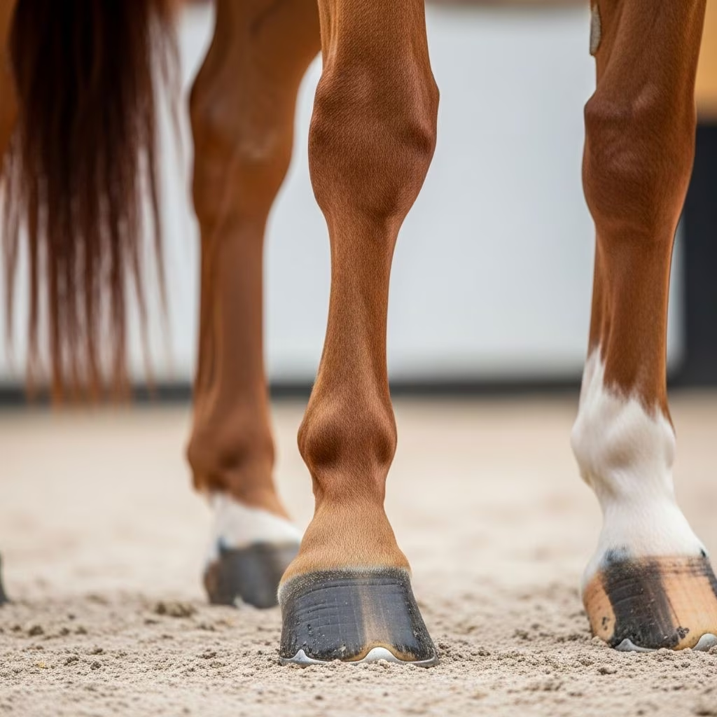
The fetlock and pastern regions represent critical components of a horse’s lower limb, enduring immense forces during locomotion. These areas frequently suffer injuries that compromise mobility and career longevity, particularly in athletic equines. This article delves into their structure, prevalent pathologies, evaluation protocols, therapeutic interventions, and proactive measures to safeguard equine soundness.
Fundamental Anatomy of the Equine Lower Limb
Grasping the intricate design of the fetlock and pastern is foundational for recognizing dysfunction. The fetlock joint, a hinge-like structure, links the cannon bone to the long pastern bone (proximal phalanx), incorporating paired proximal sesamoid bones posteriorly. This configuration facilitates flexion and extension while minimizing lateral deviations, akin to a human knuckle rather than an ankle.
Key supportive elements include the suspensory ligament, which originates from the proximal cannon bone and bifurcates into branches inserting on the sesamoids, preventing excessive fetlock drop under load. Flexor tendons—the superficial digital flexor tendon (SDFT) and deep digital flexor tendon (DDFT)—course palmarly, enabling joint flexion and shock absorption. Annular ligaments encase these tendons, maintaining alignment through the pastern.
The pastern comprises the long pastern (P1) and short pastern (P2) bones, articulating with the coffin bone distally. Their length, typically 13-17% of body length, amplifies biomechanical leverage, heightening injury susceptibility during high-speed maneuvers.
| Structure | Function | Common Vulnerability |
|---|---|---|
| Suspensory Ligament | Supports fetlock extension | Desmitis from hyperextension |
| SDFT | Flexes pastern/fetlock | Strain injuries, lacerations |
| DDFT | Supports weight-bearing | Tendinitis, core lesions |
| Sesamoid Bones | Stabilize joint posteriorly | Fractures, sesamoiditis |
Prevalent Disorders Impacting Fetlock and Pastern
Horses, especially performance breeds, face a spectrum of fetlock and pastern afflictions due to repetitive concussion and torsional stresses. Suspensory desmitis tops the list, manifesting as ligament fiber tears from overextension, often in forelimbs during jumping or racing. Inflammation yields heat, swelling, and lameness graded 2-4/5, worsening post-exercise.
Tendon strains, notably SDFT bowed tendon, arise from sudden acceleration or fatigue. The SDFT bows outward due to core fiber disruption, progressing from mild fibrillations to complete rupture. DDFT issues, though subtler, provoke deep digital flexor tenopathy with coffin joint pain. Sesamoid fractures—apical, abaxial, or basilar—stem from direct trauma or chronic overload, fracturing these sesamoids and eliciting acute, non-weight-bearing lameness.
- Sesamoiditis: Chronic inflammation erodes sesamoid margins, linked to heavy body weight or poor conformation.
- Pastern Arthritis: Degenerative joint disease erodes cartilage in pastern joints, causing stiffness and effusion.
- Annular Ligament Constriction: Fibrosis entraps flexor tendons, restricting glide and inducing digital sheath tenosynovitis.
- Fractures: P1 or P2 slab fractures from hyperextension or falls.
Hindlimb fetlocks endure unique strains from propulsion, heightening DDFT and suspensory branch vulnerabilities in disciplines like reining or cutting.
Recognizing Clinical Signs and Risk Factors
Early detection hinges on vigilant observation. Lameness localizes via flexion tests: fetlock hyperflexion reproduces pain in 80% of cases. Swelling, heat, or digital pulses signal acute inflammation; chronic cases show fibrosis or windgalls (synovial distensions).
Conformational flaws amplify risks: long toes/low heels delay breakover, overloading tendons; upright pasterns transmit concussion poorly. Overweight horses or those on hard surfaces face escalated wear. Training errors—insufficient warm-up, abrupt intensity spikes—precipitate 60% of injuries. Age factors in, with veterans prone to osteoarthritis.
Diagnostic Approaches for Accurate Assessment
Veterinary diagnostics blend clinical exams with imaging. Hoof testers and wedges isolate pastern pain; nerve blocks sequentially numb structures for pinpointing. Ultrasonography visualizes 90% of soft tissue lesions, grading fiber alignment (0-3 scale) and sheath effusions. Radiographs detect sesamoid fractures, pastern chips, or osteophytes; advanced CT/MRI delineate subtle cartilage or ligament damage.
Thermography maps heat anomalies, while gait analysis via treadmills or inertial sensors quantifies asymmetry. Synovial fluid analysis rules out sepsis in infected joints.
Treatment Modalities: From Conservative to Surgical
Management tiers by severity. Acute cases demand stall rest (4-8 weeks), ice therapy (20 min thrice daily), and NSAIDs like phenylbutazone (2-4 mg/kg BID) to curb inflammation. Supportive bandages reduce edema; therapeutic ultrasound or laser promotes healing.
Regenerative therapies shine: platelet-rich plasma (PRP) or stem cells injected ultrasonographically accelerate ligament repair, boasting 70% return-to-athlete rates. Shockwave therapy enhances vascularity in chronic desmitis. Surgical options encompass sesamoid wiring, SDFT splitting, or arthroscopic debridement for fractures/joint chips.
Rehabilitation protocols span 6-12 months: controlled hand-walking escalates to underwater treadmills, preserving fitness sans concussion. Farriery adjustments—shortening toes, adding heel support—redistribute loads effectively.
| Treatment Type | Indications | Expected Outcome |
|---|---|---|
| Rest + NSAIDs | Mild strains | 80% resolution in 4 weeks |
| PRP Injection | Core ligament tears | 65-75% performance return |
| Surgery | Fractures, ruptures | Variable; 50% athletic recovery |
| Farriery | Conformational aids | Prevents recurrence |
Rehabilitation and Long-Term Care Strategies
Post-treatment, structured rehab averts reinjury. Phase 1 (weeks 1-4): Strict box rest with daily flexion. Phase 2 (months 2-3): 10-20 min walking, escalating per ultrasound checks. Phase 3 incorporates hills, poles for proprioception. Nutritional support—omega-3s, glucosamine—bolsters tendon matrix; antioxidants combat oxidative stress.
Monitoring via serial ultrasounds ensures progressive fiber realignment before unrestricted work.
Preventive Measures for Soundness Preservation
Proactive husbandry trumps remediation. Routine farrier visits maintain balanced hooves; conditioning programs build tendon resilience gradually (10% weekly volume increase). Surfaces matter—soft footing mitigates concussion. Weight management via body condition scoring (ideal 5/9) unburdens limbs. Pre-purchase exams with flexion and imaging flag at-risk horses.
- Implement warm-ups: 15 min trot prior to intense exercise.
- Cross-train: Alternate disciplines to vary stresses.
- Bi-annual vet checks: Early ultrasound screening.
- Hoof supplements: Biotin for wall strength, indirectly aiding pastern.
Frequently Asked Questions (FAQs)
What causes most fetlock injuries in horses?
Repetitive hyperextension from galloping, jumping, or fatigue-induced flexor weakness predominantly triggers suspensory and tendon damage.
How long does recovery from suspensory desmitis take?
Mild cases resolve in 3-6 months with conservative care; severe ones require 9-12 months rehab, with 70% returning to work.
Can pastern conformation be corrected?
Farriery adjustments like wedge pads or toe extensions mitigate poor angles, though skeletal changes demand surgery in juveniles.
Is surgery always needed for sesamoid fractures?
No; conservative management suits non-displaced apical chips, while comminuted or displaced fractures necessitate pinning or removal.
How to differentiate tendon vs. ligament pain?
Ultrasound distinguishes: tendons show fusiform swelling; ligaments exhibit linear fiber gaps. Flexion worsens both, but wedges specify pastern involvement.
Prognosis and Performance Outlook
Prognoses vary: superficial SDFT tears carry 50% racing return; proximal suspensory desmitis yields 80% for pleasure horses but 40% elite athletes. Early intervention and compliance dictate success. Owners should weigh economics—$5,000-$20,000 per case—against utility.
Maintaining fetlock and pastern integrity demands holistic vigilance, blending science, skill, and stewardship for equine partners’ enduring vitality.
References
- Equine Joint Anatomy & Common Conditions — Mad Barn. 2023. https://madbarn.ca/horse-joint-anatomy/
- Large Animal Surgery: Tendon and Ligament Anatomy — University of Minnesota Libraries Publishing. 2022-10-15. https://open.lib.umn.edu/largeanimalsurgery/chapter/tendon-anatomy/
- Assessing Pastern Conformation & Its Effects — American Farriers Journal. 2021. https://www.americanfarriers.com/articles/15239-assessing-pastern-conformation-and-its-effects
- Bones, Tendons & Ligaments of the Lower Leg — Horses Inside Out. 2023. https://www.horsesinsideout.com/post/taking-the-mystery-out-of-equine-anatomy-part-3-the-bones-tendons-ligaments-of-the-lower-leg
- Guide to the Fetlock — Cryochaps. 2024. https://www.cryochaps.co.uk/guide-to-the-fetlock/
- Beyond the Hoof: Understanding the Fetlock in Horses — Oreate AI. 2023. https://www.oreateai.com/blog/beyond-the-hoof-understanding-the-fetlock-in-horses/d4fe236b1b10c5a9cab92d3bf3a9444c



