

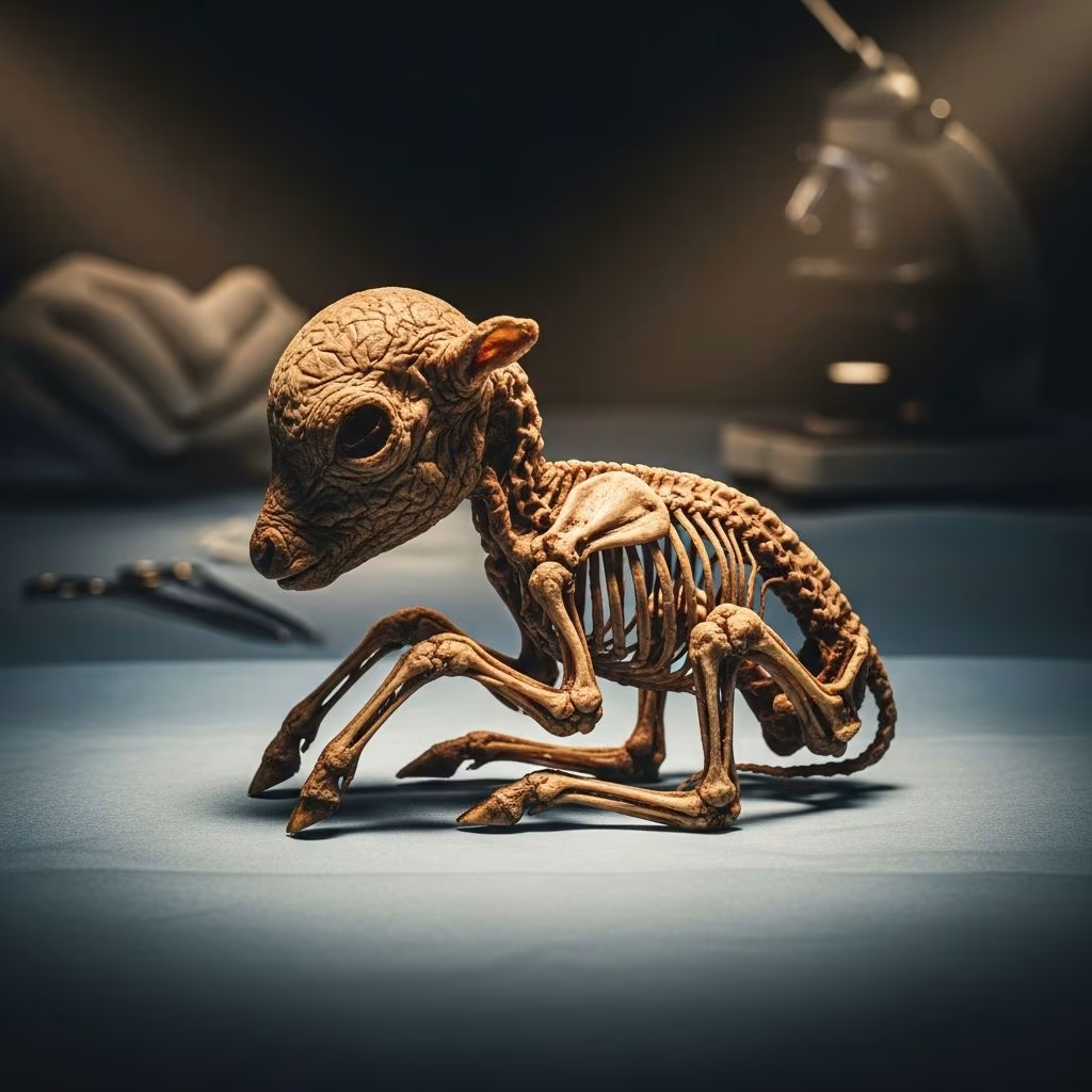
Fetal mummification occurs when a developing fetus dies in utero and the body dehydrates due to resorption of fluids, often leading to prolonged gestation in cattle and sheep as the corpus luteum persists without luteolysis.
Understanding the Biological Process
In ruminants like cattle and sheep, pregnancy maintenance relies on progesterone from the corpus luteum (CL). When fetal death happens typically between 3-8 months of gestation, the lack of a living fetus prevents the release of prostaglandins from the endometrium, allowing the CL to remain active indefinitely. This results in an abnormal pregnancy state where the dam appears pregnant but carries a non-viable, mummified fetus—a shrunken, leathery mass without fluid or odor.
The process begins with fetal demise from various triggers, followed by autolysis and dehydration via maternal circulation. Unlike abortions where fresh or autolyzed fetuses are expelled, mummification retains the remains, masking the loss and extending gestation beyond normal terms (about 280 days for cattle, 147 days for sheep).
Primary Causes in Cattle
Cattle experience fetal mummification sporadically, often linked to infectious agents or non-infectious factors. Key contributors include:
- Viral infections: Bovine viral diarrhea (BVD) disrupts early placental development.
- Parasitic invasions: Neospora caninum, detected via DNA in mummified tissues, causes mid-to-late term losses and can infect up to 35% of dairy herds endemically.
- Bacterial pathogens: Trichomoniasis and leptospirosis compromise fetal viability.
- Non-infectious issues: Umbilical cord torsion, uterine twists, genetic defects, or hormonal imbalances.
These factors halt fetal growth, initiating mummification without immediate maternal signs, prolonging the pregnancy.
Factors Triggering Mummification in Sheep
Sheep show higher mummification rates than cattle, predominantly from infectious diseases rather than mechanical issues. Unlike cattle, mummified fetuses in sheep are often expelled spontaneously. Major etiologies encompass:
- Toxoplasmosis: Toxoplasma gondii from cat feces ingestion leads to mummification at any stage, with white foci on placentomes; prime suspect for mummified cases.
- Bacterial clusters: Chlamydophila, Coxiella burnetii, and border disease virus (ToxChBCox group), causing sporadic, asymptomatic infections.
- Viral threats: Bluetongue virus induces mummification alongside stillbirths and malformations; Cache Valley virus via insect vectors prompts winter abortions.
- Nutritional deficits: Protein/energy shortages around days 90-120 of gestation exacerbate risks.
In sheep, infections often occur without flock-wide outbreaks, complicating prevalence tracking due to infrequent diagnostics.
Recognizing Clinical Indicators
Maternal signs are subtle in both species. Cattle dams remain bright and continue lactating if previously producing, with no estrus, discharge, or systemic illness. Palpation reveals a firm, irregular uterus housing a solid fetal mass sans fluid; absent uterine artery fremitus confirms death. Ultrasonography visualizes the dry, contracted fetus.
In sheep, abdominal ballotment detects the hard mass, aided by transabdominal ultrasound. Ewes may abort mummified remains spontaneously, sometimes with mild, unnoticed fever post-infection.
| Aspect | Cattle | Sheep |
|---|---|---|
| Fetal Retention | Prolonged, indefinite | Often expelled |
| Maternal Signs | None prominent | Mild fever possible |
| Diagnosis | Rectal palpation/US | Abdominal palpation/US |
| Prevalence | Uncommon | Higher incidence |
Diagnostic Approaches
Confirming mummification requires integrated exams:
- Physical assessment: Rectal (cattle) or abdominal (sheep) palpation for fluidless uterus and mummified mass.
- Imaging: Ultrasound detects absent heartbeat, fluid, or viability markers.
- Lab tests: Serology, PCR for pathogens like Neospora or Toxoplasma on fetal/placental samples; histopathology shows dehydrated tissues.
- History review: No estrus post-expected term, prior infections in herd.
Differential diagnoses include pyometra or schistosomus reflexus, ruled out via imaging and culture.
Treatment and Management Strategies
Intervention terminates the abnormal state promptly:
- Prostaglandin therapy: Single IM PGF2α injection induces luteolysis in cattle, expelling the mummy 48 hours later via manual or spontaneous vaginal passage.
- Sheep protocols: Similar hormonal induction if retained; culling common economically.
- Supportive care: Antibiotics if infection suspected; monitor for metritis post-expulsion.
Post-treatment, dams typically cycle normally, though infertility risks persist if underlying issues unaddressed.
Prevention Through Herd Health
Proactive measures curb incidence:
- Vaccination: Against BVD, bluetongue, and clostridials.
- Biosecurity: Rodent/cat control for Toxoplasma; insect netting for vectors.
- Nutrition: Balanced rations, especially mid-gestation.
- Screening: Serology for Neospora-endemic herds; cull carriers.
- Breeding hygiene: Test bulls for trichomoniasis; quarantine newcomers.
Early pregnancy checks via ultrasound identify at-risk cases.
Economic Implications for Producers
Mummification erodes profitability: one affected cow delays calving by months, costing $500+ in lost milk/meat. Flock-level outbreaks amplify losses via reduced lambing rates. Diagnostic investment (e.g., necropsies) prevents recurrence, yielding ROI through sustained fertility.
Research Insights and Future Directions
Studies highlight infectious dominance in sheep vs. multifactorial cattle cases. PCR advancements aid precise etiologies, informing targeted vaccines. Nutritional trials underscore mid-gestation feeding impacts.
Frequently Asked Questions (FAQs)
What causes the corpus luteum to persist in mummification?
No viable fetus or inflammation triggers prostaglandin release, maintaining progesterone.
Can mummified dams breed successfully afterward?
Yes, post-PGF2α treatment, most resume normal cycles unless chronic issues exist.
How do you differentiate mummification from abortion?
Mummification lacks discharge/estrus; palpation shows dry uterus vs. fluid in live pregnancies.
Is fetal mummification herd-wide?
Rarely; usually sporadic, but endemic pathogens like Neospora affect multiples.
Should mummified fetuses be submitted for testing?
Yes, for PCR/serology to identify causes and guide prevention.
References
- Fetal mummification in the major domestic species — PMC – NIH. 2018-07-26. https://pmc.ncbi.nlm.nih.gov/articles/PMC6067784/
- Detection of Neospora caninum DNA in a mummified bovine fetus — Texas A&M Veterinary Medical Diagnostic Laboratory. Undated. https://tvmdl.tamu.edu/case-studies/detection-of-neospora-caninum-dna-in-a-mummified-bovine-fetus/
- Prolonged Gestation Associated with Fetal Mummification in Cattle and Sheep — Merck Veterinary Manual. Undated. https://www.merckvetmanual.com/reproductive-system/prolonged-gestation-in-cattle-and-sheep/prolonged-gestation-associated-with-fetal-mummification-in-cattle-and-sheep
- Abortion in Sheep — MSD Veterinary Manual. Undated. https://www.msdvetmanual.com/reproductive-system/abortion-in-large-animals/abortion-in-sheep
- Infectious Causes of Abortions in Sheep — Cornell University Animal Health Diagnostic Center. Undated. https://www.vet.cornell.edu/animal-health-diagnostic-center/programs/nyschap/modules-documents/infectious-causes-abortions-sheep



