

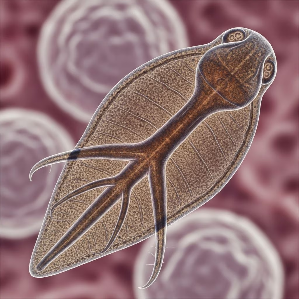
Fasciola gigantica, a significant parasitic trematode, primarily infects the livers of ruminants such as cattle, sheep, and goats, leading to fasciolosis—a condition that impairs animal health and productivity worldwide, especially in tropical and subtropical areas.
Biological Characteristics of the Parasite
This liver fluke stands out due to its impressive size, reaching lengths of up to 75 mm and widths around 15 mm, distinguishing it from its relative Fasciola hepatica, which is smaller at about 30 mm by 15 mm. Both species belong to the family Fasciolidae and share similar flattened, leaf-like bodies covered in spines, but F. gigantica predominates in warmer climates across Africa, Asia, and the Middle East. Adult worms reside in the bile ducts, where they feed on blood and tissue, causing chronic damage over time.
The parasite’s cone-shaped anterior end aids in its attachment within host tissues. Its robust morphology allows survival in harsh environments, contributing to its prevalence in wetland regions favored by its snail hosts.
Complex Lifecycle and Transmission Pathways
The lifecycle of F. gigantica is intricate, involving aquatic snails as intermediate hosts and culminating in infection of definitive mammalian hosts. Eggs, passed immature in the feces of infected animals, embryonate in freshwater within roughly two weeks, hatching into free-swimming miracidia. These miracidia seek out specific snail species, penetrating their tissues to develop through sporocyst, redia, and cercaria stages.
Cercariae emerge from snails and encyst on submerged vegetation or water surfaces as metacercariae—the infectious stage. Ruminants ingest these cysts while grazing on contaminated pastures or drinking from infested water sources. Inside the host’s small intestine, metacercariae excyst, penetrate the gut wall, traverse the peritoneal cavity, and burrow into the liver parenchyma. Maturation into egg-laying adults in the bile ducts takes approximately 3-4 months, or slightly longer for F. gigantica compared to F. hepatica.
- Egg stage: Excreted in feces, requires water to develop.
- Miracidium: Infects snails within hours of hatching.
- Intra-snail development: Multiple generations lead to cercariae release.
- Metacercariae: Survive weeks to months on plants.
- Juvenile migration: Causes acute tissue damage en route to bile ducts.
Environmental factors like temperature, humidity, and flooding enhance transmission, particularly in irrigated farmlands and marshy areas.
Geographic Distribution and Risk Factors
F. gigantica thrives in tropical zones, with high prevalence in sub-Saharan Africa, Southeast Asia, and parts of the Middle East, where livestock management intersects with snail habitats. Unlike F. hepatica’s temperate stronghold, F. gigantica favors warmer conditions, overlapping in some hybrid zones. Ruminants like buffalo and cattle serve as primary reservoirs, with sheep and goats also susceptible.
Risk escalates in regions with poor drainage, monsoon flooding, or rice paddies. Movement of infected animals spreads the parasite, while climate change may expand suitable habitats northward.
Pathophysiology: From Migration to Chronic Infestation
Infection progresses in two phases. The acute migratory phase sees juvenile flukes tunneling through liver tissue, provoking hemorrhage, inflammation, and necrosis. Hosts experience toxic reactions, fever, and eosinophilia as the body responds to larval invasion.
During the chronic biliary phase, mature flukes anchor in ducts, eroding epithelium with suckers and ingesting hepatocytes and blood. This leads to hyperplasia, fibrosis, cholangitis, and biliary obstruction. Heavy burdens exacerbate anemia, hypoalbuminemia, and weight loss due to malabsorption and chronic inflammation.
| Phase | Key Pathological Changes | Duration |
|---|---|---|
| Acute (Migratory) | Hepatic tracks, hemorrhage, inflammation | 4-8 weeks |
| Chronic (Biliary) | Duct dilation, fibrosis, anemia | Months to years |
Clinical Manifestations in Affected Livestock
Symptoms vary by burden, host species, and nutritional status. Acute fasciolosis manifests as lethargy, anorexia, abdominal pain, and anemia, often with sudden deaths in young stock. Sheep may show more severe signs than cattle due to smaller size.
Chronic cases feature emaciation, bottle-jaw edema, submandibular swelling, and reduced milk or wool production. Cattle might appear normal externally but suffer liver condemnation at slaughter, incurring economic losses. Co-infections with bacteria worsen outcomes, leading to black disease or bacillary hemoglobinuria.
Diagnostic Approaches for Field and Lab Confirmation
Detection combines history, clinical signs, and lab tests. Fecal sedimentation or flotation reveals operculated eggs (140-180 µm by 70-90 µm), though immature infections evade this method until patency at 10-12 weeks post-infection.
Serology via ELISA detects antibodies early, while coproantigen tests identify circulating antigens. Ultrasonography visualizes liver lesions, and PCR offers species-specific identification. Post-mortem, gross exams show enlarged, fibrotic livers with flukes in ducts.
- Fecal egg count: Simple, quantitative for chronic cases.
- Serological assays: Sensitive for prepatent detection.
- Imaging: Non-invasive for live animals.
- Histopathology: Confirms migration tracts.
Treatment Options and Pharmacological Interventions
Triclabendazole remains the gold standard, effective against juvenile and adult stages at 10-12 mg/kg orally, achieving over 90% efficacy. Alternatives include closantel, nitroxynil, or oxyclozanide for mature flukes, though resistance emerges in some areas. Timing treatments around peak transmission (e.g., post-flooding) maximizes impact.
Dose carefully by weight; repeat if egg counts persist. Supportive care with anthelmintics, vitamins, and improved nutrition aids recovery.
Prevention and Control Strategies for Herds
Integrated management targets the lifecycle at multiple points. Pasture drainage, fencing off wetlands, and snail control with molluscicides reduce metacercariae. Promote grazing rotation and avoid wet-season herbage.
Vaccines are experimental; focus on quarantine, strategic deworming, and monitoring. Breeding resistant stock shows promise in some breeds.
Economic and Zoonotic Implications
Fasciolosis costs billions annually in lost productivity, liver condemnations, and treatments, hitting smallholders hardest. As a zoonosis, humans risk infection from contaminated water plants, emphasizing One Health approaches.
FAQs
What animals are most affected by Fasciola gigantica?
Cattle, sheep, goats, and buffalo in tropical regions are primary hosts.
How long does it take for eggs to appear in feces after infection?
Typically 10-16 weeks, after flukes mature in bile ducts.
Is Fasciola gigantica treatable in livestock?
Yes, triclabendazole is highly effective; consult a vet for dosing.
Can humans get Fasciola gigantica from infected animals?
Yes, via metacercariae on aquatic plants, though rare in ruminant-focused areas.
What environmental conditions favor F. gigantica spread?
Warm, wet climates with abundant snails and flooded pastures.
References
- DPDx – Fascioliasis — Centers for Disease Control and Prevention (CDC). 2023. https://www.cdc.gov/dpdx/fascioliasis/index.html
- Overview of Fasciolosis — ProBiologists. 2024-10-01. https://www.probiologists.com/public/assets/articles/article-pdf-1764396648-912.pdf
- Fasciolosis in Ruminants — Semantic Scholar. 2015. https://pdfs.semanticscholar.org/ba39/121ca1cd7bd2be1d8ff7fef0b0c919a4859f.pdf
- Fascioliasis – StatPearls — National Center for Biotechnology Information (NCBI). 2023-07-17. https://www.ncbi.nlm.nih.gov/books/NBK537032/
- A review of our current understanding of parasite survival in silage — PubMed Central (PMC). 2019. https://pmc.ncbi.nlm.nih.gov/articles/PMC6582591/



