

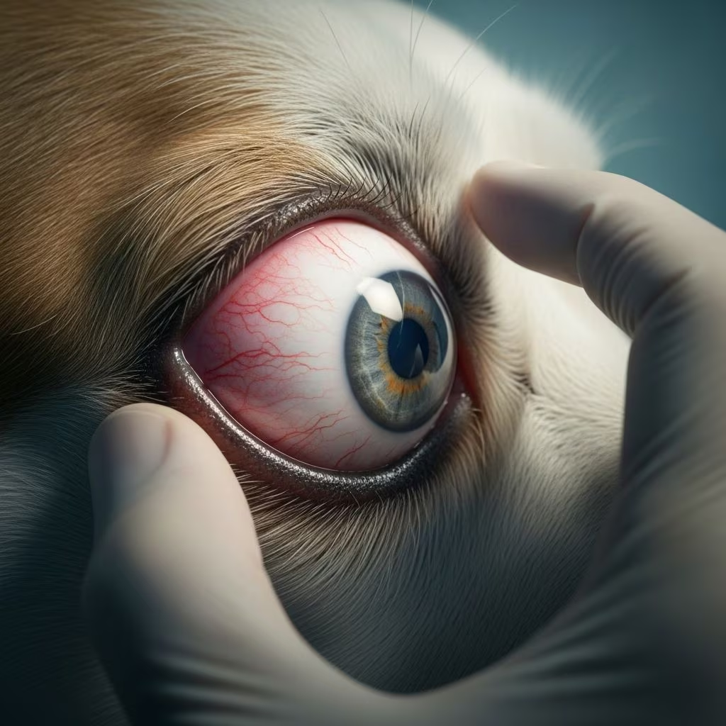
Eye prolapse represents one of the most serious ocular emergencies in veterinary medicine, affecting both dogs and cats with varying degrees of severity. When an animal’s eyeball becomes displaced forward from its normal position within the eye socket, immediate veterinary intervention becomes critical to preserve vision and prevent permanent damage. This condition encompasses several related problems, including prolapse of the third eyelid gland—commonly referred to as “cherry eye”—as well as complete forward displacement of the entire eyeball. Understanding the distinction between these presentations, recognizing early warning signs, and knowing appropriate emergency response protocols can significantly influence outcomes for affected animals.
Understanding the Anatomy of Eye Prolapse
The canine and feline eye represents a complex anatomical structure held in place by delicate connective tissues, muscles, and ligaments. The eyeball sits within the orbital socket, protected and positioned by seven extraocular muscles that control movement and a series of elastic connective tissues that maintain its forward position. Additionally, the third eyelid contains a small gland responsible for tear production—this specialized structure can become problematic when connective tissues weaken.
The third eyelid gland, scientifically termed the nictitating membrane gland, produces approximately 30 to 50 percent of an animal’s tears, making it crucial for ocular surface health and lubrication. When the fibrous attachment anchoring this gland to the eye’s inner rim deteriorates, the gland can slip forward from its normal hidden position, becoming visible at the eye’s leading edge. This condition differs from complete eye prolapse, where the entire eyeball protrudes forward—a more severe emergency requiring immediate surgical intervention.
Breed Predisposition and Risk Factors
Certain dog breeds demonstrate significantly higher susceptibility to eye prolapse conditions due to inherited structural weaknesses. Brachycephalic breeds—those with naturally flat faces such as Bulldogs, Boston Terriers, Pugs, and French Bulldogs—face elevated risk due to their distinctive eye socket anatomy. Additionally, toy and sporting breeds including Cocker Spaniels, Beagles, Chihuahuas, Shih Tzus, Lhasa Apsos, and Bloodhounds frequently develop third eyelid gland problems.
Large breed dogs such as Mastiffs, Neopolitan Mastiffs, and English Bulldogs demonstrate both higher initial incidence rates and elevated recurrence risks following surgical repair. Cats, while less commonly affected than dogs, can experience these conditions, with Burmese and Persian breeds showing particular vulnerability. Young animals, typically those under five years of age, represent the primary demographic for initial presentation, though the condition can manifest at any age depending on underlying causative factors.
Identifying Early Warning Signs and Symptoms
Pet owners and veterinary professionals should maintain vigilance for several clinical indicators suggesting eye prolapse:
- Appearance of a red or pink mass at the inner corner of the eye
- Visible protrusion beneath the third eyelid
- Excessive eye discharge or mucoid material
- Conjunctival redness and inflammation
- Squinting or signs of ocular discomfort
- Excessive tearing or dry eye symptoms
- Pawing or rubbing at the affected eye
- Light sensitivity or photophobia
- Swelling of tissues surrounding the eye
In cases of complete eye prolapse—the most severe presentation—the entire eyeball becomes displaced forward, creating an obviously distressed appearance. Animals may display severe pain, inability to close the eyelids properly, and dramatic swelling. This represents a true ophthalmologic emergency requiring immediate professional intervention within hours rather than days.
Understanding Underlying Causes
Eye prolapse in animals results from multiple interconnected factors rather than a single causative mechanism. Chronic low-grade inflammation of immune tissues within the third eyelid can lead to gland enlargement, which stretches and weakens the elastic ligaments maintaining normal position. Environmental allergen exposure, particularly in young animals encountering new allergens for the first time, frequently triggers this inflammatory cascade.
Genetic predisposition plays a substantial role, with certain breeds inheriting naturally weaker connective tissue structures around the eye. These hereditary factors combine with environmental triggers to produce clinical disease. Occasionally, trauma to the eye area can precipitate prolapse by directly damaging the supporting structures, though this occurs less frequently than genetically mediated cases.
Chronic irritation from other ocular conditions, persistent inflammation, or immune system dysregulation may contribute to the progressive weakening of connective tissues. The condition often begins intermittently, with the gland temporarily protruding before spontaneously repositioning, before eventually becoming permanently displaced.
Diagnostic Procedures and Examination
Veterinary ophthalmologists employ comprehensive examination techniques to assess eye prolapse severity and determine appropriate treatment approaches. Visual inspection provides initial assessment, revealing the characteristic red or pink mass associated with third eyelid gland prolapse. For complete eye prolapse cases, the diagnosis becomes immediately obvious, though determining whether the eye remains viable requires careful evaluation.
Ophthalmic testing may include assessment of tear production using Schirmer tear tests, evaluation of corneal reflexes and light responses, and examination of anterior chamber clarity. Photography and measurement of prolapse magnitude help guide treatment decisions. Advanced imaging such as ultrasound or CT scanning occasionally becomes necessary when assessing concurrent orbital diseases or determining whether severe trauma has caused irreversible damage.
Breed, age, and examination findings collectively inform treatment recommendations. Young animals with recent prolapse typically demonstrate excellent candidacy for surgical repair, while older animals or those with chronically prolapsed glands may require modified surgical approaches.
Immediate Emergency Management
When eye prolapse occurs, immediate protective care becomes critical before reaching veterinary facilities. For third eyelid gland prolapse, gentle cleansing with sterile saline solution helps prevent infection and reduces inflammation. Pet owners should avoid attempting to push the gland back into position without professional guidance, as improper manipulation can cause further damage.
For complete eye prolapse—the most severe scenario—applying cool sterile saline-soaked compresses helps maintain tissue viability while preventing desiccation. Prompt transport to emergency veterinary facilities becomes essential, as the window for successful eye preservation typically extends only several hours. Animals should receive pain management and sedation as appropriate while awaiting professional intervention.
Pre-operative medical therapy may include topical anti-inflammatory medications, antibiotic ointments, and systemic antibiotics to address inflammation and prevent infection. These measures prepare tissues for surgical intervention and improve healing potential.
Surgical Treatment Options and Techniques
Surgical replacement represents the gold standard treatment for eye prolapse in animals, particularly third eyelid gland prolapse. The procedure typically involves creating a small tissue pocket—termed a conjunctival fornix—in which the gland is positioned and secured with absorbable sutures. Modern surgical techniques ensure the gland remains functional while remaining properly anchored.
Multiple surgical approaches exist, with “pocketing” and “anchoring” techniques representing the most commonly employed methods. Surgeons select specific techniques based on gland size, tissue condition, breed characteristics, and individual patient factors. Successful surgical outcomes depend on precise anatomical positioning and secure tissue attachment that accommodates normal gland function.
Complete eye prolapse requires different surgical considerations. If the eye has been prolapsed for brief periods and remains viable, repositioning within the orbit becomes possible, followed by surgical securing of the orbital ligaments and supporting tissues. In cases where irreversible damage has occurred, enucleation—complete eye removal—may become necessary to prevent chronic pain and infection.
Surgical success rates for third eyelid gland prolapse repair generally exceed 90 percent when performed promptly on young animals, with nearly 100 percent success achievable in animals presenting within days of onset. Older animals with chronically prolapsed glands or those presenting months to years after initial prolapse demonstrate lower success rates and greater difficulty in repair.
Post-Operative Care and Recovery
Recovery from eye prolapse surgery typically spans three to six weeks, during which careful post-operative management significantly influences outcomes. Continued topical medications—including antibiotic and anti-inflammatory eye drops—support healing and reduce inflammation of the conjunctiva and gland tissue.
Animals require activity restriction during the healing period to prevent disruption of surgical repairs. Elizabethan collars prevent self-trauma from excessive eye rubbing or scratching. Regular veterinary follow-up examinations monitor healing progression, assess tear production, and identify any complications early.
The most common post-operative complication involves re-prolapse of the tear gland, requiring second surgical intervention. Certain breeds, particularly English Bulldogs and Neopolitan Mastiffs, demonstrate elevated recurrence risks, sometimes necessitating combined surgical techniques to achieve lasting gland repositioning.
Long-Term Consequences of Untreated Prolapse
Leaving eye prolapse unaddressed carries serious long-term ocular consequences. When the third eyelid gland remains prolapsed for extended periods, the exposed tissue becomes increasingly inflamed, infected, and desiccated. The gland’s exposed surface fails to produce tears normally, contributing to overall tear film deficiency.
Keratoconjunctivitis sicca—dry eye disease—represents the most serious long-term complication, potentially developing months or years after chronic gland prolapse. Without adequate tear production, the corneal surface becomes vulnerable to ulceration, scarring, and secondary infections. These progressive changes can ultimately result in vision loss or even blindness if not managed aggressively.
Corneal vascularization and mineralization occur as the eye attempts to heal itself in response to chronic irritation. Conjunctival tissue contact with the abnormally positioned gland causes ongoing mechanical irritation. Additionally, complete removal of a prolapsed gland—once a common but now discouraged practice—virtually guarantees future dry eye disease development.
Why Gland Preservation Matters
The tear film maintains ocular surface health through multiple mechanisms including lubrication, antimicrobial protection, and corneal nourishment. The third eyelid gland’s contribution of 30 to 50 percent of basal tear production makes it irreplaceable. Removal of this gland eliminates this tear source permanently, creating a permanent predisposition to dry eye disease.
Modern surgical techniques focus exclusively on gland preservation and repositioning rather than removal. The slight additional surgical complexity becomes worthwhile when compared against the permanent consequences of gland removal. Even when surgical repair proves technically challenging, preservation attempts remain preferable to permanent gland loss.
Prevention and Monitoring Strategies
While genetic predisposition cannot be prevented, owners of high-risk breeds should maintain heightened awareness of early warning signs. Regular ophthalmic examinations help identify intermittent prolapse before permanent displacement occurs. Minimizing environmental allergen exposure and managing allergic disease reduces inflammatory triggers.
Animals with a history of third eyelid gland prolapse—whether surgically repaired or not—require lifelong monitoring for dry eye disease development. Regular tear production testing and ophthalmic examinations become essential components of preventive care. Some animals benefit from prophylactic treatment of the contralateral eye, as many eventually develop bilateral disease.
Responsible breeding practices within predisposed breeds should include selection against animals demonstrating hereditary eye problems. Genetic counseling for breeders helps reduce disease prevalence in future generations.
Frequently Asked Questions About Eye Prolapse
Can eye prolapse resolve without surgery?
While third eyelid gland prolapse may occasionally regress temporarily, permanent resolution without surgical intervention occurs rarely. Most animals require surgical repositioning to achieve lasting correction and prevent complications.
What happens if I delay treating eye prolapse?
Delaying treatment increases risks of gland inflammation, infection, and permanent damage to the third eyelid cartilage. Extended prolapse also complicates surgical repair, potentially reducing success rates and requiring more complex surgical techniques.
Can both eyes be affected?
Yes, approximately half of animals eventually develop bilateral disease. Many ophthalmologists recommend prophylactic treatment of the unaffected eye based on breed and individual risk factors.
What is the success rate for surgical repair?
Success rates exceed 90 percent overall, with nearly 100 percent success in young animals treated promptly. Recurrence risk varies by breed, with Bulldogs and Mastiffs requiring second surgeries more frequently.
Will my pet develop dry eye disease?
Animals with prolapsed glands demonstrate elevated dry eye risk. Gland-preserving surgical repair minimizes this risk compared to gland removal, but lifelong monitoring remains important.
References
- Cherry Eye / Prolapsed Nictitans Gland Treatment — Animal Vision Center of Virginia. https://animalvisioncenterva.com/library/prolapsed-nictitans-gland-and-replacement/
- Third Eyelid Gland Prolapse (Cherry Eye) — Animal Vision Care. https://animalvisioncare.com/eye-conditions/canine/third-eyelid-gland-prolapse-cherry-eye/
- Cherry Eye in Dogs: Signs, Symptoms & Treatment Options — UVS Online. https://www.uvsonline.com/cherry-eye-prolapsed-third-eyelid-gland/
- Cherry Eye in Dogs: Signs, Symptoms, Treatments — American Kennel Club. https://www.akc.org/expert-advice/health/cherry-eye-in-dogs/
- Third Eyelid Gland Prolapse in Dogs — MSPCA-Angell. https://www.mspca.org/angell_services/third-eyelid-gland-prolapse-dogs/
- Cherry Eye in Dogs — VCA Animal Hospitals. https://vcahospitals.com/know-your-pet/cherry-eye-in-dogs
- Conditions of the Third Eyelid Factsheet — Vet Specialists UK. https://www.vetspecialists.co.uk/fact-sheets-post/conditions-of-the-third-eyelid/



