

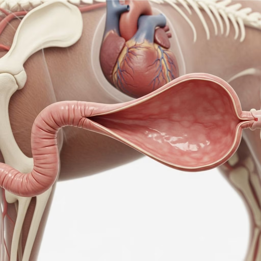
Esophageal dilatation, often linked to strictures in dogs and cats, represents a significant challenge in veterinary gastroenterology, where the esophagus widens abnormally due to impaired motility or obstructions, leading to regurgitation and nutritional deficits. This condition demands prompt intervention to restore normal swallowing and prevent life-threatening complications like aspiration pneumonia.
Understanding the Esophagus and Its Vulnerabilities
The esophagus serves as a muscular tube transporting food from the mouth to the stomach through coordinated peristaltic waves. In small animals, disruptions such as inflammation or scarring can cause segmental dilatation proximal to narrowed areas, resulting in food retention and secondary issues. Common in breeds with congenital predispositions or post-surgical cases, this pathology affects quality of life profoundly.
Primary Causes Leading to Dilatation
- Esophagitis from Reflux: Gastroesophageal reflux during anesthesia, particularly in spay surgeries, irritates the distal esophagus, fostering scar tissue formation.
- Foreign Bodies: Stuck objects cause trauma, inflammation, and subsequent narrowing with proximal pouching.
- Vomiting and Medications: Repeated emesis or oral antibiotics heighten risk, especially in females and cats with trichobezoars.
- Idiopathic or Neuromuscular: Less common, but motility disorders contribute to generalized dilatation.
These triggers often culminate in benign esophageal strictures (BES), with most cases clustering caudal to the heart base in the thoracic esophagus.
Recognizing Clinical Signs in Pets
Owners frequently notice immediate post-anesthesia regurgitation of undigested food, weight loss, and hypersalivation. Chronic cases exhibit ptyalism, halitosis, and respiratory distress from aspirated material. Unlike megaesophagus, which is diffuse, dilatation here is typically focal, proximal to a stricture, prompting urgent veterinary evaluation.
| Symptom | Mild Cases | Severe Cases |
|---|---|---|
| Regurgitation | Occasional, post-meal | Frequent, immediate |
| Weight Loss | Minimal (<10%) | Rapid (>15%) |
| Respiratory Signs | Rare | Coughing, pneumonia |
| Appetite | Normal | Anorexia |
Diagnostic Approaches for Accurate Identification
Initial assessment includes thoracic radiographs revealing dilated esophagus rostral to a stricture or foreign body shadows. Contrast studies with barium highlight narrowing and dilation dynamics, though liquid barium risks aspiration—opt for thickened mixtures. Definitive diagnosis relies on esophagoscopy, visualizing inflammation, strictures, and biopsy sites to rule out neoplasia. Cytology and histopathology differentiate benign from malignant lesions.
Treatment Strategies: Mechanical and Medical
Treatment centers on dilation therapies combined with esophagitis control. Mechanical dilation addresses strictures directly, while supportive care prevents recurrence.
Mechanical Dilation Techniques
Endoscope-Tip Dilation: Suitable for wider strictures, the endoscope advances through under visualization, stretching the lesion gradually. Effective in 75% of cases with good outcomes after 1-3 sessions.
Balloon Catheter Dilation: Preferred for narrow strictures, a deflated balloon passes via endoscope, inflates with saline to radial force, minimizing shear trauma. Sessions occur every 2-3 days, 2-4 times typically, under fluoroscopy for precision.
- Procedure steps: Anesthesia, endoscopic stricture location, balloon positioning, graded inflation, post-dilation mucosa check.
- Advanced option: Balloon-esophageal tube (BE-tube) for at-home daily dilation post-initial procedure.
Medical Management Post-Dilation
Prevent esophagitis flare-ups with:
- Proton pump inhibitors or H2 blockers (e.g., cimetidine 10 mg/kg q8h).
- Sucralfate slurry for mucosal protection.
- Promotility agents like cisapride (1 mg/kg q12h).
- Prednisone tapered course for inflammation.
- Antibiotics if infection suspected.
Nutritional support via esophagostomy or gastrostomy tubes ensures healing without oral intake.
| Method | Best For | Success Rate | Risks |
|---|---|---|---|
| Endoscope Tip | Wider strictures | 75% good | Perforation if severe esophagitis |
| Balloon Catheter | Narrow strictures | 80-90% | Tear, requires multiple sessions |
| BE-Tube | Recurrent cases | High with compliance | Tube dislodgement, infection |
Prognosis and Long-Term Outlook
Success hinges on stricture severity, location, and intervention timing. Most achieve good function after 2-3 dilations, with earlier treatment yielding superior results. Complications like perforation (rare, <5%) or recurrence demand vigilance. Owners report pets resuming normal diets, though soft foods aid initially. Euthanasia occurs in refractory cases due to costs or poor response.
Preventive Measures for Pet Owners
- Monitor post-anesthesia for regurgitation; elevate food bowls.
- Avoid table scraps reducing foreign body risk.
- Regular deworming and grooming minimize trichobezoars in cats.
- Prompt vomiting treatment prevents reflux escalation.
FAQs on Esophageal Dilatation
What triggers esophageal dilatation most often in dogs and cats?
Anesthesia-related reflux, especially during ovariohysterectomy, tops the list, alongside foreign bodies and chronic vomiting.
How many dilation sessions are typically needed?
Usually 2-4, spaced 2-3 days apart, though some resolve after one and others require more.
Is balloon dilation safe for my pet?
Yes, when guided endoscopically; risks like perforation are low (<5%), outweighed by benefits.
Can my pet eat normally after treatment?
Most do, starting with soft foods; full recovery enables balanced diets, preventing weight loss.
What if dilation fails?
Options include stents, surgical resection (rare), or feeding tubes; prognosis varies.
Recent Advances and Research Insights
Studies affirm balloon dilation’s efficacy in 18 dogs and 10 cats (median age 4 years), with repeat procedures optimizing lumen diameter. Fluoroscopy enhances precision, reducing complications. Ongoing research explores novel stents and pharmacotherapies for fibrosis prevention.
References
- Benign esophageal stricture in the dog and cat — PMC – NIH. 2000-01-01. https://pmc.ncbi.nlm.nih.gov/articles/PMC226983/
- Esophageal strictures in cats and dogs: Signs, causes and treatment — dvm360. N/A. https://www.dvm360.com/view/esophageal-strictures-cats-and-dogs-signs-causes-and-treatment
- Esophageal Balloon Dilation in Dogs — WagWalking. N/A. https://wagwalking.com/treatment/esophageal-balloon-dilation
- Balloon Dilation Of Benign Esophageal Stricture — Texas Vet Specialty. N/A. https://txvetspecialty.com/services/internal-medicine/balloon-dilation-of-benign-esophageal-stricture/
- Esophageal Stricture Management — Texas A&M Veterinary Medical Teaching Hospital. N/A. https://vethospital.tamu.edu/small-animal/internal-medicine/esophageal-stricture-management/
- Endoscopic Balloon Dilation of Benign Esophageal Strictures — Wiley Online Library. 2001-01-01. https://onlinelibrary.wiley.com/doi/abs/10.1111/j.1939-1676.2001.tb01589.x



