

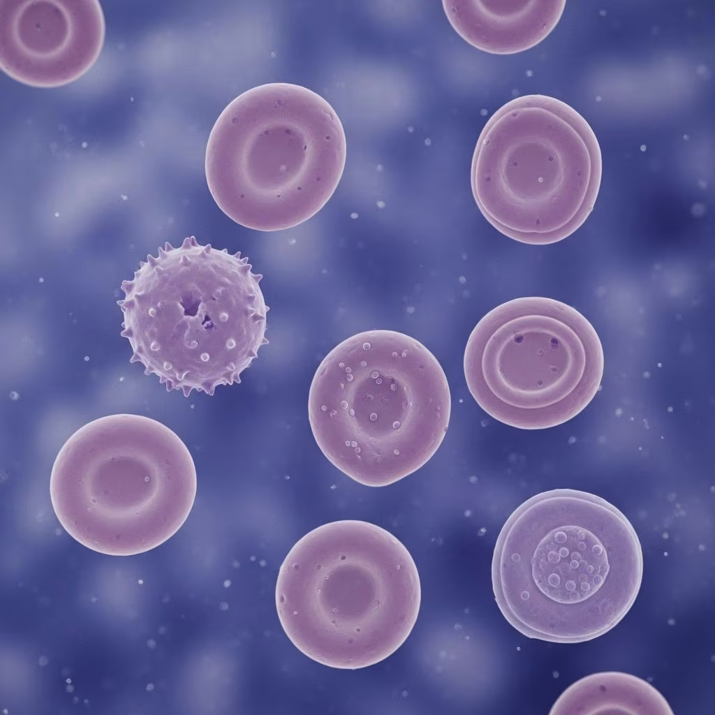
Red blood cells, or erythrocytes, serve as the cornerstone of oxygen transport in horses, ensuring muscles and organs receive vital energy during rest or intense activity. These specialized cells, packed with hemoglobin, adapt uniquely to the demands of equine physiology, but disruptions can lead to fatigue, poor performance, or life-threatening conditions.
The Anatomy of Horse Erythrocytes
Horse red blood cells stand out due to their **biconcave disc shape**, which maximizes surface area for gas exchange while allowing flexibility to squeeze through narrow capillaries. Unlike human cells, equine erythrocytes lack a nucleus and mitochondria, relying solely on glycolysis for energy to maintain shape and ion balance.
Hemoglobin, the iron-rich protein within these cells, binds oxygen in the lungs and releases it to tissues. This protein imparts the characteristic red color and enables efficient oxygen delivery under high metabolic demands, such as racing or endurance work. The biconcave form also facilitates deformation, crucial for horses with large body masses requiring robust circulation.
| Property | Horse RBCs | Human RBCs (Comparison) |
|---|---|---|
| Shape | Biconcave disc | Biconcave disc |
| Nucleus | Absent | Absent |
| Mean Corpuscular Volume (MCV) | Smaller, higher MCHC | Larger |
| Diameter | ~5-6 μm | ~7-8 μm |
This table highlights key differences, with horse cells showing higher mean corpuscular hemoglobin concentration (MCHC) for compact oxygen carrying.
How Red Blood Cells Are Produced in Horses
Erythropoiesis, the process of red blood cell formation, originates in the bone marrow from hematopoietic stem cells. These multipotent cells differentiate into committed erythroid progenitors under the influence of **erythropoietin (EPO)**, a hormone secreted by the kidneys in response to hypoxia.
When oxygen levels drop, kidneys detect this via sensors and ramp up EPO production, prompting stem cells to proliferate, mature, and release into circulation. Nutrient availability—iron, folate, vitamin B12—and cell signaling further modulate this rate. Mature equine RBCs circulate for about 140-150 days before spleen-mediated removal.
- Stem cell commitment to erythroid lineage
- Proliferation and hemoglobin synthesis
- Nucleus extrusion forming reticulocytes
- Maturation and bloodstream entry
Chronic kidney issues impair EPO output, often resulting in non-regenerative anemia. Exercise-induced hypoxia can transiently boost EPO, enhancing RBC counts in athletic horses.
Primary Functions and Metabolic Pathways
Beyond oxygen and carbon dioxide shuttling, RBCs maintain pH balance and protect against oxidative stress through metabolic pathways. Glycolysis dominates, converting glucose to ATP via insulin-independent uptake, powering Na+/K+ pumps for membrane integrity.
The pentose phosphate pathway generates NADPH to combat oxidation, preserving hemoglobin from damage. Deficiencies in enzymes like pyruvate kinase disrupt ATP production, shortening cell lifespan and causing hemolytic anemia.
In horses, high glycolytic reliance supports endurance, but stressors like intense exercise elevate oxidative demands, testing these protective mechanisms.
Common Imbalances: Anemia in Horses
**Anemia** arises when RBC counts or hemoglobin drop below normal, reducing oxygen capacity and manifesting as lethargy, pale gums, rapid breathing, and exercise intolerance. Causes span production failures, blood loss, or accelerated destruction.
- Regenerative anemia: Bone marrow ramps up output post-hemorrhage or hemolysis, detectable via increased reticulocytes (though scarce in circulation) and RDW.
- Non-regenerative: Stem cell deficits, nutrient lacks, or EPO suppression from kidney disease.
Diagnosis involves CBC showing low PCV, MCV variations, and smear exams for regeneration signs like polychromasia.
Polycythemia: Excess Red Blood Cells
Conversely, **polycythemia** features elevated RBC mass, thickening blood (increased viscosity) and straining the heart. Symptoms include sluggishness, brick-red mucosa, and thromboembolism risk.
Relative polycythemia stems from dehydration concentrating cells; absolute forms link to chronic hypoxia (e.g., lung disease) spurring EPO overproduction. Management focuses on hydration, addressing primaries, and phlebotomy in severe cases.
Aging and Removal of Equine RBCs
Horse RBCs age over ~145 days, densifying via membrane loss and hemoglobin oxidation. Density fractionation reveals low (L), medium (M), and high (H) density populations: H cells show band 3 reduction, hyper-oxidation (elevated autofluorescence), and altered Ca2+ distribution.
Senescent cells bear removal signals, phagocytosed by spleen macrophages. Hemoglobin breaks to bilirubin, iron recycled to marrow. Excess hemolysis elevates bilirubin, signaling disorders.

Advanced Markers of RBC Health and Regeneration
Detecting regeneration challenges equine vets due to minimal circulating reticulocytes. Emerging markers include RDW elevation, MCV shifts, intra-RBC Ca2+ levels, band 3 abundance via eosin-5-maleimide, and autofluorescence for oxidation.
H fraction cells exhibit high Ca2+-loaded vesicles, thiol oxidation, distinguishing them from younger L/M cells. Creatine levels and flow cytometry aid non-invasive monitoring, vital for performance horses.
Nutritional and Environmental Influences
Iron, copper, and cobalt support hemoglobin synthesis; deficiencies mimic anemia. Pasture management prevents geophagia-induced overloads. Dehydration from transport or heat stress spuriously elevates PCV, masking true status.
Antioxidants like vitamin E mitigate exercise-induced hemolysis in racehorses.
Veterinary Diagnostics and Monitoring
Routine CBC tracks PCV (32-52%), RBC count (6-12 x10^6/μL), hemoglobin (11-19 g/dL). Advanced: flow cytometry for density/Ca2+, bone marrow biopsy for production assessment.
| Test | Normal Range (Horses) | Low Indicates | High Indicates |
|---|---|---|---|
| PCV | 32-52% | Anemia | Polycythemia/Dehydration |
| RBC Count | 6-12 x10^6/μL | Production failure | Overproduction |
| RDW | Low variability | – | Regeneration |
Management Strategies for Owners
Prevent via balanced diet, parasite control, regular deworming to curb blood loss. For anemic horses: address causes (e.g., supplements for iron-deficiency), EPO mimics under vet guidance. Monitor athletes post-exertion for oxidative hemolysis.
Frequently Asked Questions (FAQs)
What causes pale gums in my horse?
Pale mucous membranes signal anemia from low RBCs/hemoglobin, often due to parasites, blood loss, or nutrition gaps. Vet CBC confirms.
How long do horse red blood cells live?
Approximately 140-150 days, removed by spleen after senescence markers appear.
Can exercise improve RBC counts?
Yes, via hypoxia-induced EPO, boosting production in trained horses.
Is polycythemia dangerous?
Yes, blood thickening risks clots and heart strain; hydrate and treat underlying issues.
What diet supports healthy RBCs?
Iron-rich forages, copper-balanced feeds; avoid excesses to prevent hemochromatosis.
References
- Red Blood Cells in Horses – Equine Research Database — Mad Barn. 2023. https://madbarn.com/research-topics/red-blood-cells/page/3/
- Red Blood Cells of Horses – Horse Owners — Merck Veterinary Manual. 2023-10-01. https://www.merckvetmanual.com/horse-owners/blood-disorders-of-horses/red-blood-cells-of-horses
- Aging Markers in Equine Red Blood Cells — PMC (Peer-reviewed). 2019-08-15. https://pmc.ncbi.nlm.nih.gov/articles/PMC6650539/
- Red Blood Cells in Animals – Circulatory System — MSD Veterinary Manual. 2024. https://www.msdvetmanual.com/circulatory-system/hematopoietic-system-introduction/red-blood-cells-in-animals
- Erythrocyte Structure and Function — Wiley Online Library (Schalm’s Veterinary Hematology). 2023. https://onlinelibrary.wiley.com/doi/abs/10.1002/9781119500537.ch20



