

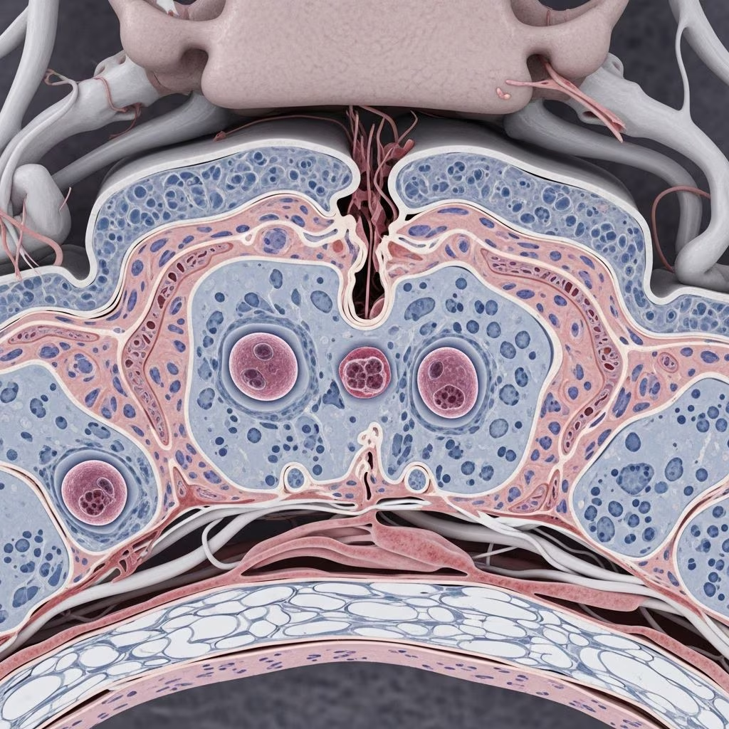
Equine Protozoal Myeloencephalitis (EPM) stands as one of the most prevalent neurological conditions affecting horses in the Americas. This disease arises from protozoan parasites invading the central nervous system, leading to a spectrum of debilitating symptoms that can severely impact a horse’s quality of life and performance capabilities. Horse owners, trainers, and veterinarians must recognize the nuances of EPM to facilitate early intervention and improve outcomes.
The Biological Basis of EPM
At its core, EPM results from infection by Sarcocystis neurona, the primary culprit, with Neospora hughesi playing a less frequent role. These protozoa target the brain, spinal cord, and associated nerves, causing inflammation and tissue damage. Horses serve as dead-end hosts, meaning they cannot propagate the parasite’s life cycle, but they suffer the consequences of its presence.
The transmission pathway hinges on opossums, the definitive hosts for S. neurona. These marsupials shed infectious sporocysts in their feces, which contaminate pastures, hay, feed, or water sources. Horses inadvertently ingest these sporocysts while grazing or eating. Once inside the horse, the parasites breach the blood-brain barrier, establishing residence in neural tissues and inciting an immune response that exacerbates damage.
Recognizing the Signs: From Subtle to Severe
EPM’s clinical presentation is notoriously variable, often mimicking musculoskeletal injuries or other neurological disorders. Symptoms can emerge gradually or abruptly, affecting any part of the central nervous system. This unpredictability complicates initial assessments.
- Asymmetric ataxia: Uncoordinated movements, more pronounced on one side, particularly in hind limbs. Horses may stumble, weave, or widen their stance for balance.
- Muscle weakness and atrophy: Progressive loss of muscle mass, often focal, in areas like the hindquarters, topline, or face. Affected horses struggle with slopes or head elevation.
- Cranial nerve dysfunction: Facial drooping, head tilting, difficulty swallowing (dysphagia), ear droop, or abnormal eye positioning.
- Behavioral and cerebral changes: Lethargy, obtundation, seizures, or altered consciousness in cases involving the brain.
- Other indicators: Unusual lameness, loss of sensation, excessive sweating, or collapsing.
Horses under stress—such as during transport, intense training, or concurrent illness—are particularly vulnerable, with symptoms potentially escalating rapidly. Notably, infected animals rarely show fever or pain, distinguishing EPM from infectious or traumatic conditions.
Factors Influencing Disease Development
Not all exposed horses develop clinical EPM; seroprevalence exceeds 50% in some U.S. regions, yet overt disease is less common. Immune competence plays a pivotal role—robust systems may suppress the parasite asymptomatically for years. Stressors like weaning, racing, or immunosuppression trigger clinical manifestation.
| Risk Factor | Description | Impact on Susceptibility |
|---|---|---|
| Geographic Location | Areas with opossum populations (Americas) | High exposure risk |
| Stress Levels | Training, travel, illness | Triggers onset |
| Age | Young or performance horses | Increased incidence |
| Feed Contamination | Hay/silage with feces | Direct transmission vector |
| Immune Status | Compromised immunity | Rapid progression |
Diagnostic Approaches for Accurate Identification
Diagnosing EPM demands a multifaceted strategy due to overlapping symptoms with conditions like equine motor neuron disease (EMND), herpesvirus myeloencephalopathy, or laminitis. Veterinary evaluation begins with a thorough neurologic exam assessing gait, proprioception, and reflexes.
Key diagnostics include:
- Serology: Serum:CSF ratios via ELISA or IFAT to detect antibodies, indicating CNS involvement if CSF titers exceed serum.
- PCR Testing: Detects parasite DNA in CSF, though sensitivity varies.
- Imaging and Exclusion: Rule out trauma, abscesses via myelography or MRI when available.
Classic signs like asymmetric gait deficits and focal atrophy heighten suspicion, prompting confirmatory tests.
Treatment Protocols: Halting Progression
Early, aggressive therapy yields the best prognosis, with many horses achieving partial or full recovery. Antiprotozoal medications form the cornerstone:
- Ponazuril or Diclazuril: Oral formulations targeting protozoan replication, administered for 28 days.
- Sulfadiazine/Pyrimethamine: Combination therapy, often with folinic acid to mitigate bone marrow suppression.
- Supportive Care: Anti-inflammatories (e.g., NSAIDs), vitamin E for neuroprotection, physical therapy.
Treatment success hinges on lesion location and chronicity—spinal cases respond better than brainstem involvement. Relapses occur in 10-30% of cases, necessitating re-treatment.
Prevention Strategies: Breaking the Transmission Chain
Proactive measures minimize exposure:
- Secure feed storage to deter opossums.
- Remove wildlife attractants from barns.
- Fence pastures excluding opossums.
- Hay disinfection via heating (though not fully validated).
- Stress reduction and immune support via balanced nutrition.
Vaccines remain unavailable, underscoring environmental control’s importance.
Prognosis and Long-Term Management
With prompt treatment, 60-80% of horses improve significantly, though full athletic return varies. Chronic cases may require retirement. Monitoring post-treatment via re-exams prevents relapse.
Frequently Asked Questions (FAQs)
What causes EPM in horses?
EPM stems primarily from Sarcocystis neurona ingested via opossum-contaminated feed or water.
Is EPM contagious between horses?
No, horses are dead-end hosts and cannot transmit it directly.
How long does EPM treatment last?
Typically 4 weeks, with potential extensions for non-responders.
Can EPM be fatal?
Untreated severe cases can lead to recumbency and euthanasia, but treatment improves survival.
How can I prevent EPM on my farm?
Eliminate opossum access to feed and water sources.
Emerging Research and Future Directions
Ongoing studies refine diagnostics, like advanced PCR and biomarkers, while exploring novel therapies. Consensus guidelines from veterinary bodies emphasize multimodal approaches.
References
- Equine Protozoal Myeloencephalitis — Equine Disease Communication Center. Accessed 2026. https://www.equinediseasecc.org/EPM
- Equine Protozoal Myeloencephalitis — Michigan State University College of Veterinary Medicine. Accessed 2026. https://cvm.msu.edu/hospital/services/equine-services/for-owners/equine-protozoal-myeloencephalitis
- Equine Protozoal Myeloencephalitis – Nervous System — Merck Veterinary Manual. Accessed 2026. https://www.merckvetmanual.com/nervous-system/equine-protozoal-myeloencephalitis/equine-protozoal-myeloencephalitis
- Equine Protozoal Myeloencephalitis: An Updated Consensus — PMC (PubMed Central). 2016-06-28. https://pmc.ncbi.nlm.nih.gov/articles/PMC4913613/
- Equine Protozoal Myeloencephalitis (EPM) — UC Davis Center for Equine Health. Accessed 2026. https://ceh.vetmed.ucdavis.edu/health-topics/equine-protozoal-myeloencephalitis-epm
- Equine Protozoal Myeloencephalitis (EPM) in Horses — PetMD. Accessed 2026. https://www.petmd.com/horse/conditions/neurological/equine-protozoal-myeloencephalitis-epm-horses
- EPM and How to Prevent it — Irongate Equine Clinic. Accessed 2026. https://www.irongateequine.com/education/epm-and-how-to-prevent-it



