

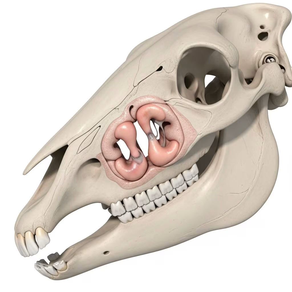
The paranasal sinuses in horses form a complex network of air-filled cavities within the skull that play key roles in respiration, head weight reduction, and voice production. These structures, including the frontal, maxillary, dorsal conchal, ventral conchal, sphenopalatine, and middle conchal sinuses, interconnect via specific channels to the nasal cavity, facilitating drainage and airflow.
Anatomy of the Equine Paranasal Sinuses
Horses possess seven paired sinus compartments, divided into rostral and caudal systems by the maxillary septum. The rostral system includes the rostral maxillary sinus (RMS) and ventral conchal sinus (VCS), while the caudal system encompasses the caudal maxillary sinus (CMS), dorsal conchal sinus (DCS), frontal sinus (FS), sphenopalatine sinus, and middle conchal sinus.
Key connections occur through sinonasal channels: the rostral sinonasal channel links to the RMS and VCS, entering via the nasomaxillary aperture in the middle nasal meatus. The caudo-lateral channel accesses the CMS directly, with fewer variations noted across specimens. The FS communicates broadly with the DCS, often termed the conchofrontal sinus due to this fusion.
- Rostral Maxillary Sinus (RMS): Houses roots of cheek teeth (e.g., 109/209), accessible via specific trephination sites.
- Caudal Maxillary Sinus (CMS): Contains roots of molars 110/210 and 111/211; links to FS via frontomaxillary opening.
- Ventral Conchal Sinus (VCS): Rostral extension, bulla-penetrable for diagnostics.
- Dorsal Conchal Sinus (DCS): Caudal part interconnects with FS.
- Frontal Sinus (FS): Large dorsal cavity, trephined medial to eyes.
Volumetric studies via computed tomography (CT) reveal symmetric channels bilaterally, crucial for surgical planning to restore physiologic drainage.
Common Causes of Sinus Conditions in Horses
Sinus disorders arise from infections, obstructions, trauma, or developmental issues. Primary sinusitis often stems from bacterial entry via teeth or nasal passages, leading to purulent accumulation. Secondary causes include cheek tooth apices penetrating sinuses, especially in younger horses where roots extend into RMS/CMS.
| Category | Examples | Prevalence Factors |
|---|---|---|
| Infectious | Bacterial (Streptococcus spp.), viral predispositions | High in stabled horses with poor ventilation |
| Obstructive | Cysts, neoplasms, ethmoidal hematomas | Progressive, often unilateral |
| Trauma | Fractures from falls or kicks | Acute onset post-injury |
| Dental | Tooth root abscesses, periodontitis | Common in 4-6 year olds |
Embryologically, sinuses develop from nasal meatus protrusions into maxillary diploe, explaining shared nasomaxillary entries vulnerable to blockages.
Recognizing Clinical Signs
Horses with sinus disease exhibit nasal discharge (unilateral purulent for infections/dental issues, bilateral for generalized), facial swelling, respiratory noise, and halitosis. Chronic cases show weight loss, depression, or foul odor. Young horses may present with dental-related empyema, while cysts cause progressive distortion.
- Unilateral mucopurulent discharge: Suspect dental or neoplastic.
- Bilateral serous: Viral or allergic.
- Facial deformity: Cysts or tumors.
- Neurologic signs: Extension to brain via sphenopalatine.
Diagnostic Approaches
Diagnosis integrates history, endoscopy, imaging, and sampling. Nasal endoscopy visualizes discharge origin and bullae integrity. Radiography assesses fluid lines, tooth roots, and masses, though superimposition limits detail.
Advanced CT provides 3D sinus mapping, channel patency, and precise volumes, essential for surgery. Ultrasound detects ventral conchal bulla thickening. Fluid cytology/radiography confirms infection vs. sterile processes.
Trephination Techniques for Sampling
Trephination creates portals for lavage, visualization, or biopsy. Sites vary by age and sinus:
- FS: 5 cm from midline, medial canthi line.
- RMS (young horses): Caudal facial crest site avoids teeth.
- CMS: Infraorbital foramen reference.
Maxillary nerve blocks prevent reaction; Galt trephines ensure stability.
Treatment Strategies
Therapy targets cause: antimicrobials for infections, surgery for obstructions. Frontal sinus trephination with flushing resolves empyema in 70-80% acute cases. Chronic disease requires sinusotomy—wavefront or cell-flap methods preserve mucosa.
Dental issues demand extraction or repulsion via sinus portals. Cysts/neoplasms need marsupialization or ablation. Postoperative care includes daily flushes, systemic antibiotics, and monitoring.
Surgical Innovations
Transnasal approaches leverage sinonasal channels for minimal invasion. CT-guided planning reconstructs physiologic pathways, improving outcomes.
Prevention and Management Tips
Regular dental checks, prompt respiratory treatment, and stable hygiene reduce risks. Early endoscopy for discharge prevents chronicity. Nutritional balance supports immunity.
Frequently Asked Questions (FAQs)
What is the most common sinus issue in horses?
Primary sinusitis, often bacterial from dental roots or infections.
How do you diagnose equine sinus cysts?
Endoscopy shows nasal mass; CT confirms size/location.
Is surgery always needed for sinusitis?
No, acute cases respond to flushing/antibiotics; chronic/obstructive require intervention.
Can sinus disease affect performance?
Yes, via airflow restriction and discomfort.
What is the prognosis post-surgery?
Excellent for infections (90%+ resolution); guarded for tumors.
Understanding equine paranasal sinuses empowers proactive care, minimizing complications from these vital structures.
References
- The sinonasal communication in the horse: examinations using computed tomography and three-dimensional reconstruction — PLoS ONE (PMC). 2014-04-08. https://pmc.ncbi.nlm.nih.gov/articles/PMC3995307/
- Paranasal sinus anatomy and trephination technique (Proceedings) — dvm360. N/A. https://www.dvm360.com/view/paranasal-sinus-anatomy-and-trephination-technique-proceedings
- Paranasal sinus disease – Large Animal Surgery — University of Minnesota Libraries Publishing. N/A. https://open.lib.umn.edu/largeanimalsurgery/chapter/paranasal-sinus-disease/
- Diseases of the Paranasal Sinuses in Horses — Merck Veterinary Manual. N/A. https://www.merckvetmanual.com/respiratory-system/respiratory-diseases-of-horses/diseases-of-the-paranasal-sinuses-in-horses



