

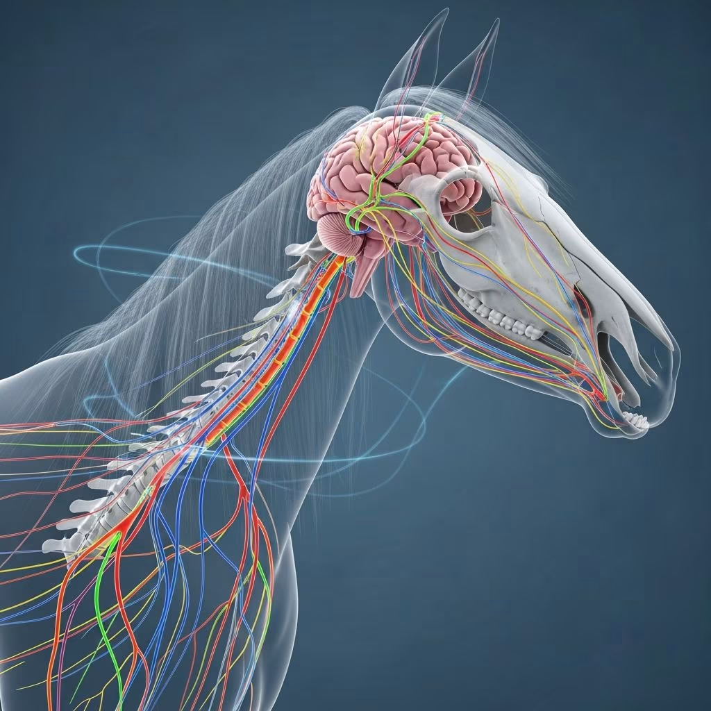
The nervous system in horses orchestrates every aspect of their physiology, from precise movements during a gallop to automatic processes like breathing. This intricate network enables horses to perceive their environment, respond to stimuli, and maintain homeostasis, making it foundational for health and performance.
Overview of the Nervous System Divisions
Horses possess a sophisticated nervous system divided into two primary components: the central nervous system (CNS) and the peripheral nervous system (PNS). The CNS, comprising the brain and spinal cord, serves as the command center, processing sensory input and issuing motor commands. The PNS extends this control outward, linking the CNS to muscles, organs, and sensory receptors throughout the body.
This division ensures seamless integration of voluntary actions, such as trotting or jumping, with involuntary ones, like digestion or heart regulation. Understanding these parts helps owners recognize signs of dysfunction early, potentially preventing severe issues like ataxia or paralysis.
Central Nervous System: Brain and Spinal Cord
The CNS forms the core of equine neurology. Protected by the skull and vertebral column, it coordinates complex behaviors and reflexes.
Brain Anatomy and Roles
The equine brain consists of several key regions, each with specialized functions. The cerebrum, the largest section, handles higher processing, including learning patterns and sensory integration. Adjacent to it, the cerebellum fine-tunes coordination, balance, and muscle tone, crucial for agile maneuvers in riding or racing.
Deeper structures like the brainstem manage vital reflexes: respiration, heartbeat, and swallowing. The midbrain acts as a relay, facilitating signal transmission and eye movement control via cranial nerves. Horses’ brains prioritize reactive instincts over abstract reasoning, enhancing survival through rapid responses.
- Cerebrum: Processes vision, hearing, and memory formation.
- Cerebellum: Ensures smooth gait and posture.
- Brainstem: Regulates autonomic life-support functions.
Spinal Cord Structure and Functions
Extending from the brainstem through the vertebral canal, the spinal cord acts as a bidirectional conduit for neural signals. Segmented into cervical, thoracic, lumbar, and sacral regions, it corresponds to body areas: cervical for neck and forelimbs, thoracic-lumbar for trunk and hindlimbs, sacral for tail and pelvis.
Local injuries yield localized symptoms; for instance, cervical damage might cause forelimb dragging, while lumbar issues affect hindquarter propulsion. The cord also mediates spinal reflexes, like the tail flick, bypassing the brain for speed.
| Spinal Region | Body Area Controlled | Common Symptoms of Damage |
|---|---|---|
| Cervical | Neck, forelimbs | Toe dragging, neck stiffness |
| Thoracic/Lumbar | Trunk, hindlimbs | Hindlimb weakness, ataxia |
| Sacral | Tail, pelvis | Tail paralysis, incontinence |
Peripheral Nervous System Components
The PNS bridges the CNS to the periphery, divided into somatic (voluntary control) and autonomic (involuntary regulation) branches. Somatic nerves handle skeletal muscle movement and sensory feedback, while autonomic governs organs.
Sensory and Motor Pathways
Sensory (afferent) nerves relay environmental data—touch, pain, temperature—to the CNS, enabling quick reactions vital for prey animals. Motor (efferent) nerves deliver commands to muscles for actions like fleeing predators.
This duality supports proprioception, allowing horses to navigate uneven terrain without visual cues. Damage here manifests as numbness or spasticity.
Cranial Nerves: Head and Sensory Specialists
Twelve pairs of cranial nerves originate from the brain, managing head-specific functions. Key examples include:
- Olfactory (I): Detects scents for foraging.
- Optic (II): Processes visual input for spatial awareness.
- Trigeminal (V): Sensory for face, motor for chewing; branches like infraorbital aid nerve blocks.
- Facial (VII): Controls expressions, ear/twitch movements.
- Vagus (X): Influences heart, gut, respiration—key for stress responses.
Issues like head tilt or dysphagia signal cranial nerve compromise, often from trauma or infection.
Autonomic Nervous System in Equines
The autonomic nervous system (ANS) operates subconsciously, balancing sympathetic (fight-or-flight) and parasympathetic (rest-and-digest) activities. Sympathetic surges boost heart rate and alertness during exertion; parasympathetic conserves energy post-exercise.
In horses, ANS dysregulation appears in colic (parasympathetic dominance slowing gut motility) or anhidrosis (sympathetic failure impairing sweat). Maintaining balance through proper management enhances resilience.
Protective Mechanisms and Meninges
The CNS is shielded by meninges: dura mater (outer tough layer), arachnoid mater (middle web-like), and pia mater (innermost adherent to tissue). Cerebrospinal fluid cushions impacts, while the blood-brain barrier filters toxins.
These defenses are critical; infections like equine protozoal myeloencephalitis breach them, causing inflammation.
Common Neurological Challenges in Horses
Horses face risks from trauma, infections, and toxins. Equine herpesvirus targets the CNS, inducing ataxia; EPM from protozoa erodes coordination.
Symptoms include circling, muscle tremors, or knuckling. Early veterinary intervention via CSF analysis or imaging improves outcomes. Preventive vaccination and biosecurity are paramount.
Diagnostic Approaches for Nerve Issues
Vets employ neurological exams assessing gait, cranial nerve function, and reflexes. Tail pull tests spinal integrity; menace response checks vision.
Advanced tools like myelography visualize cord compression. Bloodwork rules out metabolic causes.
Maintaining Nervous System Health
Nutrition supports myelin sheath integrity; vitamin E deficiency risks degeneration. Exercise promotes neuroplasticity, while deworming prevents migrating larvae damage.
Stable management minimizes trauma risks, like slips on wet floors.
FAQs
What causes wobbler syndrome in horses?
Wobblers stem from cervical vertebral instability, compressing the spinal cord and causing hindlimb ataxia. Surgical stabilization often resolves it.
How do cranial nerves affect horse performance?
They enable precise head movements, vision, and swallowing, directly impacting trailering tolerance and feeding efficiency.
Can stress damage a horse’s nervous system?
Chronic stress disrupts ANS balance, potentially leading to ulcers or immune suppression, indirectly affecting nerves.
What is the role of reflexes in equine neurology?
Spinal reflexes like the panniculus (skin twitch) confirm intact peripheral pathways, aiding injury localization.
How does age impact the horse’s nervous system?
Older horses risk neurodegeneration, manifesting as senile ataxia; antioxidants may mitigate oxidative damage.
References
- The Equine Nervous System — Horse Education Online. 2023. https://www.horseeducationonline.com/post/the-equine-nervous-system
- Head – CVM Large Animal Anatomy — University of Minnesota Pressbooks. 2024. https://pressbooks.umn.edu/largeanimalanatomy/chapter/neck-head/
- 14.5: Parts of the Nervous System — Medicine LibreTexts (University of Minnesota). 2023. https://med.libretexts.org/Bookshelves/Veterinary_Medicine/Anatomy_and_Physiology_of_Animals_(Lawson)/14:_Nervous_System/14.05:_Parts_of_the_Nervous_System
- Understanding the Horse’s Brain: Anatomy, Function & Related … — Mad Barn (Veterinary Nutrition). 2024. https://madbarn.com/horse-brain-anatomy/
- Horse Anatomy — PetMD (Veterinary Resource). 2025. https://www.petmd.com/horse/horse-anatomy
- The Nervous System — Equine Medical Service (.edu affiliate). 2023. https://emsvet.com/the-nervous-system/



