

Movement dysfunction in horses represents one of the most frequently encountered challenges in equine veterinary medicine, affecting animals across all disciplines and age groups. When a horse displays abnormal gait patterns or demonstrates reluctance to bear weight on a limb, the underlying etiology may range from superficial soft tissue injuries to complex osseous pathology. Prompt and accurate diagnosis is paramount, as delayed intervention can result in secondary compensatory injuries, prolonged recovery periods, and compromised athletic performance. This comprehensive overview examines the clinical evaluation process that equine veterinarians employ to systematically identify the anatomical and physiological origins of locomotor dysfunction.
Understanding the Scope of Movement Abnormalities
Locomotor dysfunction in equines encompasses a broad spectrum of clinical presentations stemming from multiple pathophysiological mechanisms. Horses experiencing movement impairment typically present with pain localized to musculoskeletal structures, mechanical impediments that restrict normal joint and limb movement, or neurological conditions affecting motor function or sensory perception. The distinction between acute and chronic presentations significantly influences diagnostic strategy and therapeutic planning.
Acute-onset movement dysfunction typically manifests suddenly, frequently following trauma, overexertion, or acute injury. A horse may become acutely reluctant to bear weight following a penetrating wound to the foot, such as a nail puncture, or may develop severe gait abnormality following a misstep or direct impact injury. Conversely, chronic locomotor dysfunction develops insidiously over extended periods, often progressing gradually as underlying degenerative or inflammatory processes advance. Conditions such as osteoarthritis, chronic tendon strain, or progressive ligamentous pathology exemplify this presentation pattern.
Initial Clinical Consultation and History Gathering
The diagnostic process commences with a comprehensive history-taking session that provides essential context for the physical examination. The veterinarian obtains detailed information regarding the temporal onset of the movement dysfunction, factors that exacerbate or ameliorate the condition, prior episodes of similar complaints, and the horse’s work history and performance demands. Environmental factors, recent changes in footing or training intensity, and previous injuries also factor into the differential diagnosis formulation.
Understanding the horse’s discipline and performance expectations guides diagnostic prioritization. Racehorses demonstrate predilections for certain conditions—such as carpus (knee) arthritis—related to the biomechanical stresses inherent to high-speed galloping. Dressage and advanced performance horses frequently present with hindlimb suspensory apparatus disorders stemming from collection demands and repetitive flexion stresses. Trail and pleasure horses may present with different injury patterns reflecting their activity profiles.
Physical Examination at Stationary Position
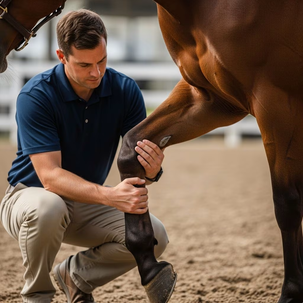
The examination of the stationary equine patient forms the foundation of the diagnostic assessment. The veterinarian performs a systematic evaluation of conformation, postural stance, and structural alignment. Visual inspection identifies obvious asymmetries, swelling, heat, or wounds that may localize the dysfunction to specific regions.
Through careful palpation—using trained hands to detect subtle temperature variations, edema, and abnormal tissue texture—the veterinarian systematically examines each limb segment. Specific attention focuses on identifying areas of heat associated with inflammation, soft tissue swelling suggesting fluid accumulation or traumatic edema, and localized pain responses upon direct pressure or manipulation. The hoof receives particular scrutiny, as hoof-related pathology represents the most prevalent cause of locomotor dysfunction in horses.
Hoof testers—specialized instruments that apply graduated pressure to specific hoof regions—help identify painful areas within the foot structure. Responses to hoof test pressure often indicate subsolar pathology, abscessation, or deep foot structures sensitive to compression. Careful assessment of foot balance, shoe condition, and any evidence of previous therapeutic shoeing modifications provides additional diagnostic clues.
Dynamic Assessment During Locomotion
Evaluation of the moving horse provides essential diagnostic information unavailable from static examination alone. The veterinarian observes the horse during multiple gaits and under varying circumstances to detect gait abnormalities, asymmetries, and pain-related movement modifications.
Walking and trotting in a straight line constitute fundamental components of the dynamic assessment. The veterinarian observes stride length symmetry, rhythm regularity, foot landing patterns, and weight-bearing preferences. Shortening of stride on the affected limb, asymmetrical rhythm, or reluctance to fully load a particular leg provides critical diagnostic information. The horse may demonstrate a “nodding” head motion when trotting, wherein the head elevates as the lame limb contacts the ground—a compensatory mechanism reflecting pain reduction through altered weight distribution.
Trotting on varied surfaces—including firm ground, soft footing, and slight inclines—provides dynamic information regarding pain localization and condition severity. Many horses demonstrate worsening gait abnormality on harder surfaces due to increased concussive forces, while others show improvement as softer footing reduces impact loading. Lunging in circles stresses specific limb structures asymmetrically and often exacerbates pain when lameness originates from certain anatomical regions.
Specialized Physical Examination Techniques
Flexion tests constitute valuable diagnostic tools that isolate and stress specific anatomical structures while the horse stands at rest. During a flexion test, the veterinarian manually flexes a joint or series of adjacent joints, maintaining flexion for approximately one minute, then releases the limb and observes the horse’s movement during the subsequent trot steps. Heightened pain responses or exacerbated gait abnormality following flexion tests strongly suggest that pain originates in the flexed structures.
Various flexion protocols target different anatomical regions. Lower limb flexion tests stress the foot, fetlock, and lower cannon bone structures, while proximal limb flexion tests evaluate carpal, tarsal, and higher limb structures. The response pattern—whether lameness improves, worsens, or remains unchanged—guides anatomical localization.
Manipulation and passive range-of-motion testing detect mechanical restrictions, joint instability, or pain elicited by specific movements. The veterinarian performs gentle passive flexion and extension of joints, noting any resistance, abnormal movement patterns, or pain responses that might indicate structural pathology.
Diagnostic Nerve and Joint Anesthesia
When clinical examination and flexion testing fail to definitively localize pain, diagnostic anesthetic techniques provide more precise anatomical identification. These procedures involve carefully injecting local anesthetic agents either near specific nerves (nerve blocks) or directly into synovial structures such as joints, bursae, and tendon sheaths (intrasynovial anesthesia).
The nerve block procedure employs a systematic approach, typically beginning with anesthetic injections at distal limb levels and progressing proximally if lameness persists. After each injection, the horse’s movement is reassessed at a trot. Improvement or resolution of lameness following anesthetic administration strongly indicates that pain originates in the anesthetized region. The systematic progression ensures anatomical precision and prevents unnecessary anesthetic administration.
Joint blocks and bursal anesthesia specifically target synovial structures. This technique proves particularly valuable for identifying pathology within specific joints, as the injected anesthetic directly bathes the structures of interest. Improvement in gait following intrasynovial anesthesia localizes dysfunction to that particular joint or synovial structure.
Radiographic Imaging Assessment
Once clinical examination and anesthetic localization have identified the anatomical region of pain, radiographic imaging typically constitutes the first advanced diagnostic step. Standard radiographs (X-rays) utilize ionizing radiation to create two-dimensional images of osseous structures and some soft tissues.
Radiographic evaluation identifies bone fractures, osseous remodeling associated with chronic arthritis, abnormal bone density, and periosteal reactions indicating healing processes or inflammatory disease. Orthogonal projections—images obtained from multiple angles—provide comprehensive visualization of complex three-dimensional structures.
Radiography demonstrates certain limitations. Early cartilage damage, soft tissue injuries to tendons and ligaments, and subtle bone lesions may not appear on standard radiographs. Additionally, some anatomical regions prove technically challenging to radiograph adequately, limiting diagnostic utility in certain cases.
Ultrasonographic Evaluation
Ultrasound imaging employs high-frequency sound waves to create detailed images of soft tissues, making it exceptionally valuable for evaluating tendons, ligaments, bursae, joint capsules, and other non-osseous structures. The technique produces real-time images without ionizing radiation, allowing dynamic assessment of tissue movement and structure.
Ultrasonography readily identifies tendon fiber disruption, ligamentous strain patterns, bursal effusion, and soft tissue inflammation. The comparison of affected limbs with contralateral normal limbs facilitates detection of subtle abnormalities. Serial ultrasonographic examinations over time document healing progression and identify complications such as adhesion formation or incomplete tissue repair.
Advanced Imaging Modalities
When standard radiographs and ultrasound fail to definitively establish diagnosis, advanced imaging techniques provide exceptional detail regarding complex pathology. Magnetic resonance imaging (MRI) produces detailed three-dimensional representations of both bone and soft tissues with superior soft tissue contrast resolution. MRI excels at identifying cartilage damage, ligamentous injuries, bone marrow pathology, and subtle soft tissue lesions invisible on conventional imaging.
Standing equine MRI systems allow safe imaging without general anesthesia risks, delivering diagnostic clarity in over 90% of lameness cases. Computed tomography (CT) provides excellent osseous detail through cross-sectional imaging, particularly valuable for complex tarsal and carpal pathology. Positron emission tomography (PET) identifies metabolically active regions indicating inflammation or injury, complementing structural imaging modalities.
Treatment Planning and Prognosis
Accurate diagnosis through systematic clinical evaluation and appropriate imaging establishes the foundation for rational therapeutic intervention. Treatment approaches vary substantially based on underlying cause, ranging from simple conservative management to surgical intervention. The veterinarian develops individualized treatment plans considering the horse’s age, intended use, overall health status, and client resources.
Regenerative therapies—including platelet-rich plasma injection, stem cell therapy, shock wave treatment, and intrarticular biologic preparations—complement conventional treatments in many cases. Complementary approaches such as acupuncture, therapeutic massage, and controlled rehabilitation programs support healing and restoration of function.
Frequently Asked Questions
What causes lameness in horses?
Lameness results from pain in musculoskeletal structures, mechanical movement restriction, or neurological conditions. Common causes include foot abscesses, joint arthritis, tendon or ligament injuries, bone fractures, infections, and developmental orthopedic disease. Specific breeds and disciplines show predilections for particular conditions due to biomechanical demands.
How long does lameness diagnosis typically require?
The diagnostic timeline varies based on condition complexity. Straightforward cases may resolve with initial clinical examination and radiographs within one to two visits. Complex cases requiring advanced imaging and multiple diagnostic blocks may extend over several weeks as additional information accumulates and anatomical localization progresses.
Can lameness resolve without veterinary intervention?
While some acute minor injuries may improve with rest, untreated lameness frequently progresses to secondary compensatory injury as the horse alters gait to minimize pain. Prompt diagnosis and appropriate treatment prevent unnecessary suffering and optimize recovery outcomes.
What is the difference between diagnostic blocks and treatment blocks?
Diagnostic blocks employ local anesthetic to identify pain location and confirm diagnosis. Treatment blocks utilize therapeutic agents to reduce inflammation and pain for therapeutic benefit. Diagnostic blocks guide treatment decisions, while therapeutic blocks provide direct symptom relief.
Conclusion: Systematic Approach to Diagnostic Excellence
The comprehensive evaluation of equine locomotor dysfunction represents a systematic progression from clinical observation through increasingly sophisticated diagnostic techniques. This methodical approach—beginning with history and physical examination, proceeding through dynamic assessment and anesthetic localization, and culminating in targeted imaging—ensures accurate identification of underlying pathology. Veterinary professionals employing this disciplined diagnostic framework optimize treatment outcomes, minimize secondary complications, and facilitate return to athletic function. The investment in thorough diagnostic evaluation ultimately conserves time, reduces treatment costs, and maximizes the probability of successful resolution.
References
- Lameness in Horses — PetMD. Accessed 2026. https://www.petmd.com/horse/conditions/musculoskeletal/lameness-horses
- Lameness Diagnostics and Treatment — Alpine Equine Hospital. Accessed 2026. https://alpineequine.net/lameness-diagnostics-and-treatment
- From Block to Image to Treatment: A Step-by-Step Guide to Equine Lameness Diagnosis — Hallmarq. 2025-11-06. https://hallmarq.net/2025/11/06/from-block-to-image-to-treatment-a-step-by-step-guide-to-equine-lameness-diagnosis/
- Understanding Lameness in Horses: Causes, Diagnosis — Bluegrass Equine. 2023-09-15. https://www.bluegrassequine.com/site/blog/2023/09/15/understanding-lameness-horses
- Lameness Diagnosis & Treatment in Lebanon — Lebanon Equine Clinic. Accessed 2026. https://lebanonequineclinic.com/services/lameness-diagnosis-treatment/



