

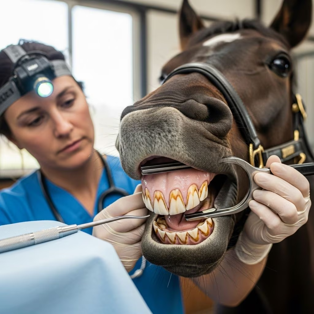
Horses possess a specialized dental system adapted for their herbivorous diet, featuring continuously erupting teeth that require regular attention to prevent discomfort and performance issues. This article delves into the anatomy, maintenance, and disorders of equine dentition, providing horse owners and veterinarians with actionable knowledge.
Unique Features of Horse Tooth Anatomy
Equine teeth are classified as hypsodont, meaning they have elongated crowns that erupt gradually throughout the horse’s life to offset wear from constant grinding of fibrous forage. Adult horses typically have 36 to 44 teeth, including incisors, canines, premolars, and molars.
The basic structure of each tooth consists of four primary tissues: the pulp at the core, surrounded by dentin, enamel forming the outer ridges, and cementum covering the reserve crown. The pulp houses nerves, blood vessels, and odontoblasts that produce dentin for protection. Dentin varies in types—primary, secondary regular, secondary irregular, and tertiary—each contributing to the tooth’s resilience against wear and injury.
Enamel, the hardest layer, creates the ridged chewing surfaces but cannot regenerate once damaged, necessitating protective positioning between dentin and cementum. Cementum encases much of the tooth, especially in cheek teeth, aiding in structural integrity.
| Tooth Tissue | Function | Key Characteristics |
|---|---|---|
| Pulp | Vital core | Nerves, blood supply, odontoblasts |
| Dentin | Protective layer | Multiple subtypes; produced ongoing |
| Enamel | Hard outer ridges | Non-regenerative; forms occlusal surface |
| Cementum | Outer covering | Abundant in reserve crowns |
Types and Arrangement of Equine Teeth
Horses have distinct tooth categories tailored to their grazing habits. The front incisors (six upper and six lower) clip grass, while canines serve males primarily for display or combat, often lacking occlusion. Cheek teeth—premolars and molars—grind food and number up to 24, with the first premolar (wolf tooth) sometimes absent or vestigial.
- Incisors: Used for cropping vegetation; feature infundibula (cups) for age estimation.
- Canines: Present in most males; lower ones positioned rostrally without contact.
- Premolars: Three per quadrant in adults; aid initial grinding.
- Molars: Three per quadrant; triangular or rectangular shapes for efficient mastication.
- Wolf Teeth: Non-erupting first premolars; often extracted if interfering with bits.
The upper jaw (maxilla) is wider than the lower (mandible) by about 23%, a condition called anisognathia, leading to natural sharp point development on upper outer and lower inner edges during lateral chewing movements.
Development and Eruption Patterns
Foals are born with deciduous teeth that shed as permanent ones emerge. Cheek teeth erupt sequentially, with the last premolar potentially misaligning if not straight, causing malocclusions. Teeth wear at 2-3 mm annually, with reserve crowns up to 9-10 cm long ensuring lifelong function.
Deciduous formula differs from permanent: foals have temporary incisors, canines, and premolars, transitioning fully by age 5. Permanent cheek teeth grow from reserve crowns embedded in bone or sinuses, especially maxillary ones extending into paranasal spaces.
Routine Dental Maintenance Practices
Regular “floating”—rasping sharp points—prevents soft tissue injuries from hooks and ramps formed due to uneven wear. Horses should receive exams at least annually, more frequently for performance animals or seniors.
Comprehensive oral exams go beyond floats, assessing occlusion, gum health, and eruption rates influenced by periodontal conditions. Sedation enables thorough visualization, including wolf tooth removal and bit seat creation for comfort.
Recognizing and Addressing Dental Abnormalities
Common issues include wave mouth (undulating occlusal plane), steps (height disparities between adjacent teeth), and ramps/hooks impeding chew cycles. These cause quidding (dropping forage), weight loss, colic risk, and behavioral changes like head tossing.
- Sharp Points: From anisognathia; cut cheeks/tongue.
- Malocclusions: From eruption errors or jaw imbalances.
- Fractures/Infundibular Necrosis: Lead to abscesses; more common in incisors.
- < periodontal Disease: Alters eruption, exacerbating waves.
Advanced tools like radiographs detect subsurface issues, while motorized floats ensure precise corrections.
Age Determination via Dental Inspection
Equine age estimation relies on incisor wear, angle changes, and infundibulum visibility. Young teeth curve convex labially; older ones flatten, forming a shallower arcade. Galvayne’s groove appearance signals advanced age.
| Age Range | Incisor Indicators |
|---|---|
| 0-1 week | Nipple teeth present |
| 6-12 months | Central incisors erupt |
| 2-5 years | Full permanent incisors; cups visible |
| 6-10 years | Cups disappear; teeth level off |
| 20+ years | Triangular shape; heavy wear |
Preventive Strategies for Long-Term Oral Health
Feed long-stem hay to promote natural grinding, minimizing grain to reduce uneven wear. Monitor for sinus issues or tumors affecting maxillary teeth. Early intervention preserves reserve crown length, vital for seniors.
Professional care by equine dentists or vets trained in occlusion is crucial, as DIY attempts risk iatrogenic damage.
FAQs on Horse Dental Health
What is floating and how often is it needed?
Floating smooths enamel points; yearly for most, biannual for athletes or weanlings.
Do all horses have wolf teeth?
No, about 30-50% do; removal advised if causing bit discomfort.
Can dental issues cause lameness or colic?
Yes, pain alters gait; poor chewing increases impaction risk.
How do I spot dental problems at home?
Look for undigested hay in manure, drooling, foul breath, or resistance to bridling.
Are senior horses’ teeth fully managed?
With proper care, yes; supplements aid weakened structures.
References
- Back to Basics: Dental Anatomy — The Horse. 2014-approx. https://thehorse.com/19389/back-to-basics-dental-anatomy/
- Dental Anatomy of Horses — Colorado State University (vivo.colostate.edu). Accessed 2026. https://vivo.colostate.edu/hbooks/pathphys/digestion/pregastric/horsepage.html
- Dental Diseases — Saginaw Valley Equine Clinic. Recent. https://saginawvalleyequine.com/experience/equine-education/dental-diseases/
- Equine Dental Anatomy: Wisconsin Equine Clinic & Hospital — YouTube/Wisconsin Equine Clinic. Recent. https://www.youtube.com/watch?v=TY7QI5P82mo
- Proceedings of the AAEP 2002 – Dentistry — IVIS/AAEP. 2002-11-01. https://www.ivis.org/sites/default/files/library/aaep/2002/910102000421.PDF
- The Equine Oral Exam: Beyond Sharp Points and Floats — Alpine Equine. Recent. https://alpineequine.net/blog/173253-the-equine-oral-exam-beyond-sharp-points-and-floats
- Dental Anatomy and Nomenclature — CABI Digital Library. 2014-approx. https://www.cabidigitallibrary.org/doi/pdf/10.5555/20143185636



